Spinal Anatomy Chart
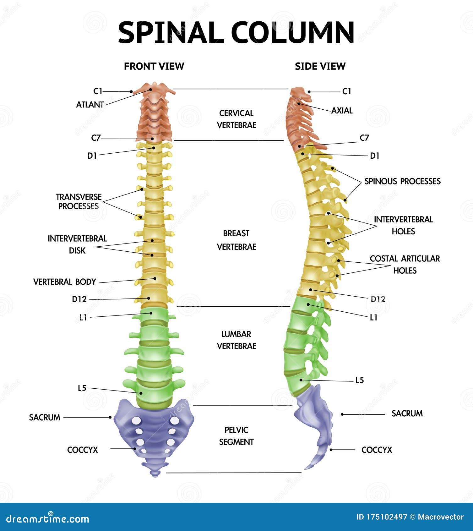
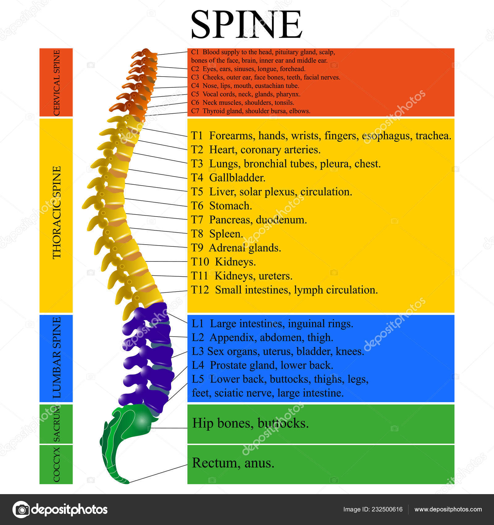
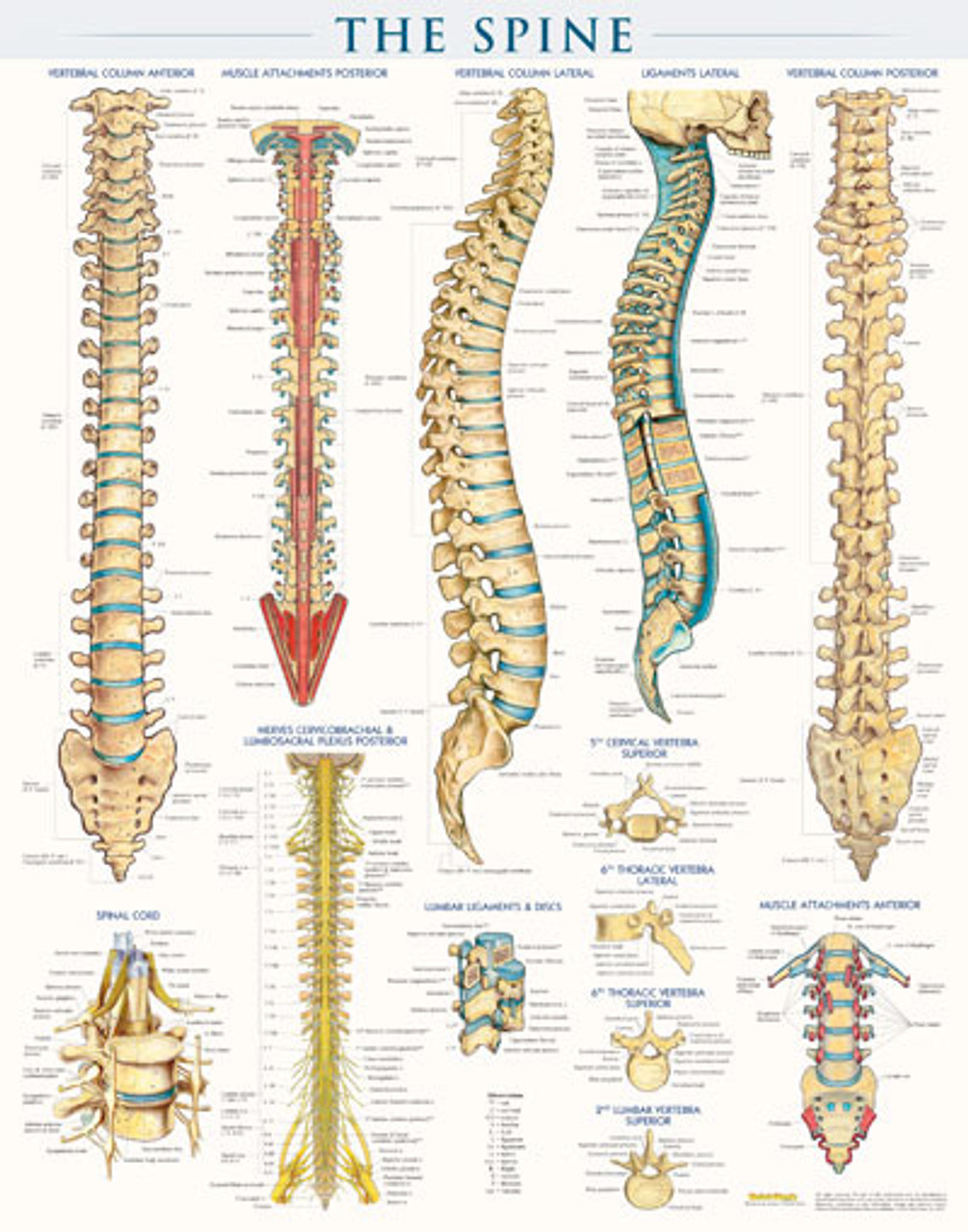
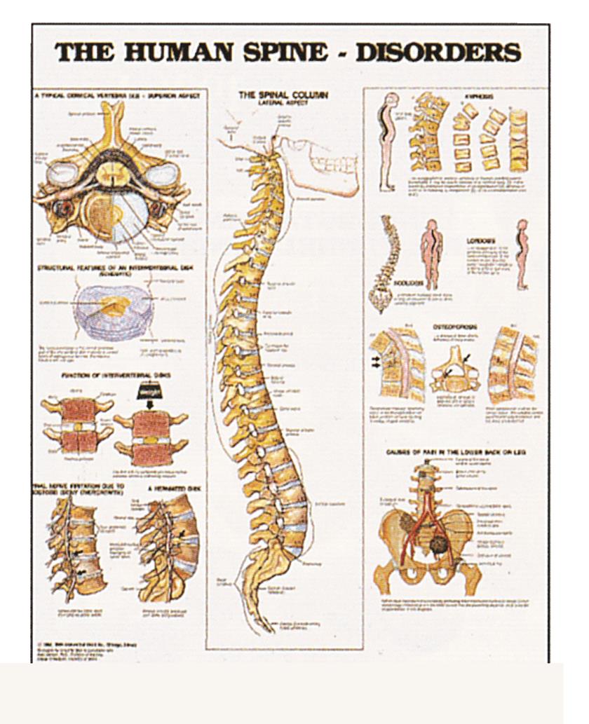
Spinal Anatomy Chart - Web your spine is made up of vertebrae (bones), disks, joints, soft tissues, nerves and your spinal cord. Exercises can strengthen the core muscles that support your spine and prevent back injuries and pain. You can see the cervical vertebrae labeled at the top, the thoracic vertebrae labeled in the middle and the lumbar vertebrae labeled towards the bottom. The spinal cord is part of the central nervous system and consists of a tightly packed column of nerve tissue that extends downwards from the brainstem through the central column of the spine. Each segment of the spinal cord provides several pairs of spinal nerves, which exit from vertebral canal through the intervertebral foramina. There are 8 pairs of cervical, 12 thoracic, 5 lumbar, 5 sacral, and 1 coccygeal. All of these bones and sections are important to the spine’s ability to function properly. Web the spine diagram below highlights all of the vertebrae labeled. Web this human anatomy module is composed of diagrams, illustrations and 3d views of the back, cervical, thoracic and lumbar spinal areas as well as the various vertebrae. Neck problems can cause neck pain and/or pain that radiates down the arms to the hands and fingers. Web the spine diagram below highlights all of the vertebrae labeled. Exercises can strengthen the core muscles that support your spine and prevent back injuries and pain. The spinal cord runs through its center. Web sacrum (sacral region) spine anatomy overview video. Cervical, thoracic, lumbar, sacral, and coccygeal. Web explore the anatomy and structure of the 26 bones that make up the spine with innerbody's 3d model. It is within this region that the nerves to the arms arise via the brachial plexus, and where the cervical plexus forms providing innervation to. It contains the osteology, arthrology and myology of the spine and back. Web the cervical portion. While many of us take the benefits. The spinal column combines strong bones, unique joints, flexible ligaments and tendons, large muscles and highly sensitive nerves. It's a delicate structure that contains nerve bundles and cells that carry messages from your brain to the rest of your body. The spinal cord begins at the base of the brain and extends into. Web sacrum (sacral region) spine anatomy overview video. Many of the nerves of the. It contains the osteology, arthrology and myology of the spine and back. Web last updated november 14, 2022 • 59 revisions •. Web it comprises the vertebral column (spine) and two compartments of back muscles; Web your spine is made up of vertebrae (bones), disks, joints, soft tissues, nerves and your spinal cord. While many of us take the benefits. Web your lumbar spine supports the upper two sections of your spine — the seven vertebrae in your neck (cervical spine) and 12 vertebrae in your chest (thoracic spine) — and the weight of your. Neck problems can cause neck pain and/or pain that radiates down the arms to the hands and fingers. While many of us take the benefits. Web spine anatomy, diagram & pictures | body maps. Web the spine (vertebral column) of a typical adult is composed of 32 vertebrae divided into five sections. What is the spinal cord? Exercises can strengthen the core muscles that support your spine and prevent back injuries and pain. It contains the osteology, arthrology and myology of the spine and back. What is the spinal cord? Neck problems can cause neck pain and/or pain that radiates down the arms to the hands and fingers. Web this article discusses the anatomy of the spine. The column can be divided into five different regions, with each region characterised by a different vertebral structure. Throughout the spine, intervertebral discs made of. Spinal pain can arise from problems in the bones, nerves, or other soft tissues. Web your spine is made up of vertebrae (bones), disks, joints, soft tissues, nerves and your spinal cord. All of these. Web the vertebral column (spine or backbone) is a curved structure composed of bony vertebrae that are interconnected by cartilaginous intervertebral discs. The spinal cord is part of the central nervous system and consists of a tightly packed column of nerve tissue that extends downwards from the brainstem through the central column of the spine. Web the cervical portion of. It's a delicate structure that contains nerve bundles and cells that carry messages from your brain to the rest of your body. Web this human anatomy module is composed of diagrams, illustrations and 3d views of the back, cervical, thoracic and lumbar spinal areas as well as the various vertebrae. Web sacrum (sacral region) spine anatomy overview video. It is. While many of us take the benefits. Web explore the anatomy and structure of the 26 bones that make up the spine with innerbody's 3d model. Web the vertebral column (spine or backbone) is a curved structure composed of bony vertebrae that are interconnected by cartilaginous intervertebral discs. Web the spine’s four sections, from top to bottom, are the cervical (neck), thoracic (abdomen,) lumbar (lower back), and sacral (toward tailbone). Each segment of the spinal cord provides several pairs of spinal nerves, which exit from vertebral canal through the intervertebral foramina. Web the spine diagram below highlights all of the vertebrae labeled. The column can be divided into five different regions, with each region characterised by a different vertebral structure. It contains the osteology, arthrology and myology of the spine and back. Web it comprises the vertebral column (spine) and two compartments of back muscles; The spinal cord begins at the base of the brain and extends into the pelvis. Mayo clinic does not endorse companies or products. Throughout the spine, intervertebral discs made of. Cervical, thoracic, lumbar, sacral, and coccygeal. Five bones in the lower back—the lumbar spine; The spinal column combines strong bones, unique joints, flexible ligaments and tendons, large muscles and highly sensitive nerves. You can see the cervical vertebrae labeled at the top, the thoracic vertebrae labeled in the middle and the lumbar vertebrae labeled towards the bottom.
Spinal Anatomy Poster 18" X 24" Clinical Charts and Supplies

Spine Anatomy Realistic Chart Stock Vector Illustration of health

Spinal Nerves Anatomical Chart Spine and Cranial Nervous System

Diagram Human Spine Name Description All Sections Vertebrae Vector

Spinal chart You Make it 1Corinthians 620 Pinterest

Spine Structure Poster Clinical Charts and Supplies

Blog Lamb Chiropractic and Wellness

Rudiger Anatomie The Human Spine Laminated Anatomy Chart

Spinal Nerve Chart Anatomy

Wall Chart The Human Spine Disorders (Single) Hillcroft Supplies
Exercises Can Strengthen The Core Muscles That Support Your Spine And Prevent Back Injuries And Pain.
12 Bones In The Chest—The Thoracic Spine;
What Is The Spinal Cord?
It Also Explores Common Conditions Affecting The Spine And When Someone May.
Related Post: