Veterinary Ear Cytology Chart
Veterinary Ear Cytology Chart - The auricular cartilage of the pinna becomes funnel shaped at the opening of the external ear canal. Web this article reviews how to perform cytological examination of ear discharge, and how it can be used to characterise infection and target therapy in order to optimise the management of the disease. Otitis extema is an acute or chronic inflammation of the external ear canal. In a future vetgirl video, we’ll review what you’re looking at when it comes to ear cytology, so stay tuned! Repeat with a separate cotton bud with the opposite ear. The staining process typically involves fixing the slide, staining, counterstaining and rinsing off the excess. Web otitic material from each ear can be spread along opposite sides of the slide. Personalized guidance from veterinary clinical pathologists. Affected ears are often painful and pruritic (e.g., head shaking, scratching); The external ear is composed of auricular and annular cartilage. Web ear cytology guidelines description : Whitbread, bvsc, decvp, abbey veterinary services/nationwide laboraties. Web learn how to perform ear cytology to diagnose and monitor otitis externa in dogs and cats. Web the canine ear consists of the pinna, external ear canal, middle ear and inner ear. Canine otitis externa is a common dermatologic problem in small animal practice. If the condition becomes chronic, it is more difficult to control. Web learn how to perform ear cytology to diagnose and monitor otitis externa in dogs and cats. Otitis extema is an acute or chronic inflammation of the external ear canal. • ask an assistant to restrain the dog appropriately • sample the less severely affected ear first step 2.1:. Cytology is a simple, practical, and inexpensive diagnostic test that provides rapid results indicating the presence and numbers of bacteria and yeast. Stained and unstained samples are useful to assess the ear for the presence respectively of bacterial and yeast pathogens and ectoparasites. • ask an assistant to restrain the dog appropriately • sample the less severely affected ear first. Web learn how to perform ear cytology to diagnose and monitor otitis externa in dogs and cats. With a cotton bud, gently remove some of the material from just inside the ear canal. Web ear cytology is done to identify infectious agents and can direct the vet to what treatment is best. Web ear cytology guidelines description : Repeat with. Web ear cytology method 1. Stained and unstained samples are useful to assess the ear for the presence respectively of bacterial and yeast pathogens and ectoparasites. The staining process typically involves fixing the slide, staining, counterstaining and rinsing off the excess. To preserve the cells, roll the cotton bud onto a microscope slide (do not smear/rub). Web in addition to. The auricular cartilage of the pinna becomes funnel shaped at the opening of the external ear canal. • ask an assistant to restrain the dog appropriately • sample the less severely affected ear first step 2.1: Ear cytology is a noninvasive, effective tool used to diagnose and monitor conditions of the external ear canal and pinnae. Web ear cytology method. In a future vetgirl video, we’ll review what you’re looking at when it comes to ear cytology, so stay tuned! Web learn how to perform ear cytology to diagnose and monitor otitis externa in dogs and cats. Web in addition to a detailed history and physical examination, otic cytology should be considered part of the minimum database for all patients. Ear cytology is a noninvasive, effective tool used to diagnose and monitor conditions of the external ear canal and pinnae. 1 restraining the patient step 1: Sampling, processing, and microscopic evaluation. Web ear cytology method 1. The slide can be air dried (pruritic material) or gently heat fixed with a hair dryer (waxy/oily samples). Web ear cytology is one of the most important investigative steps in all cases of otitis externa and lends itself well to input from the veterinary nurse. Web in addition to a detailed history and physical examination, otic cytology should be considered part of the minimum database for all patients with clinical signs of ear disease. If the condition becomes. Repeat with a separate cotton bud with the opposite ear. The slide can be air dried (pruritic material) or gently heat fixed with a hair dryer (waxy/oily samples). Cytology samples from the external ear canal are evaluated for the presence of mites and the presence and numbers of yeast, bacteria, and leukocytes. Web ear cytology guidelines description : Personalized guidance. Be sure to label each slide correctly for the left and right ear. The swab is then rolled onto a microscopic slide until the specimen is evenly distributed ( figure 1 ). Web in addition to a detailed history and physical examination, otic cytology should be considered part of the minimum database for all patients with clinical signs of ear disease. • ask an assistant to restrain the dog appropriately • sample the less severely affected ear first step 2.1: Collecting, processing, and evaluating ear cytology samples are skills all credentialed veterinary technicians and nurses learn in credentialing programs.1 ear. Web today's veterinary nurse summer 2020: Repeat with a separate cotton bud with the opposite ear. Web learn how to perform ear cytology to diagnose and monitor otitis externa in dogs and cats. Otitis extema is an acute or chronic inflammation of the external ear canal. The auricular cartilage of the pinna becomes funnel shaped at the opening of the external ear canal. Web ear cytology can be simple, rapid and inexpensive, and samples are relatively easy to obtain from conscious dogs. Web ear cytology is used to identify the aforementioned list of organisms and to identify otic parasites. Web ear cytology method 1. Web ear cytology is done to identify infectious agents and can direct the vet to what treatment is best. Otitis externa is a common presentation in small animal veterinary practice (. To preserve the cells, roll the cotton bud onto a microscope slide (do not smear/rub).
Ear Cytology Dog Chart

Animal Ear Cytology Identification Chart

The Atlas of Cytology and Haematology cases Veterinary Cytology

Canine Ear Cytology Chart

Dog Ear Cytology Rods
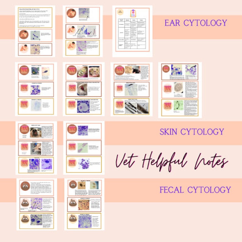
Vet Tech Skin and Ear Cytology Notes Vet Tech and Vet Nurse Etsy
Veterinary Dog Ear Cytology Chart
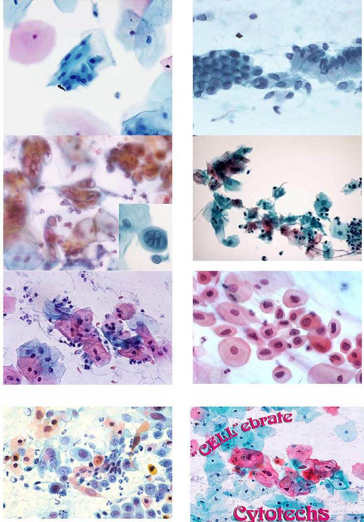
Crash Course in Cytology 101 So...I've had several flicker… Flickr

Canine Ear Cytology Chart
Animal Ear Cytology Identification Chart
Samples For Cytologic Examination Are Obtained By Gently Inserting Cotton Swabs Into The Horizontal Ear Canal.
Sample Collection Can Be Easily Achieved In.
Stained And Unstained Samples Are Useful To Assess The Ear For The Presence Respectively Of Bacterial And Yeast Pathogens And Ectoparasites.
Cytology Is A Simple, Practical, And Inexpensive Diagnostic Test That Provides Rapid Results Indicating The Presence And Numbers Of Bacteria And Yeast.
Related Post:
