Stemi Leads Chart
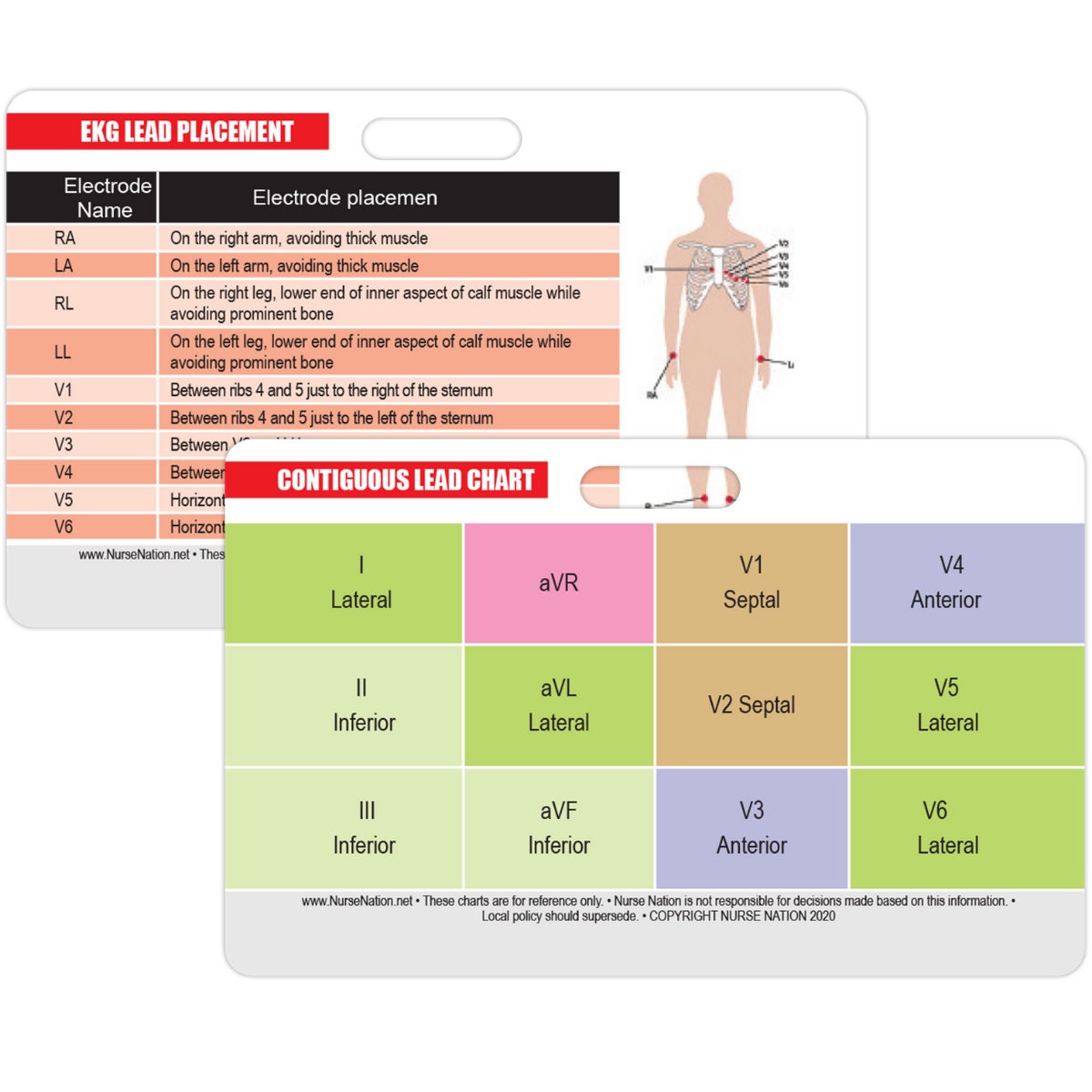
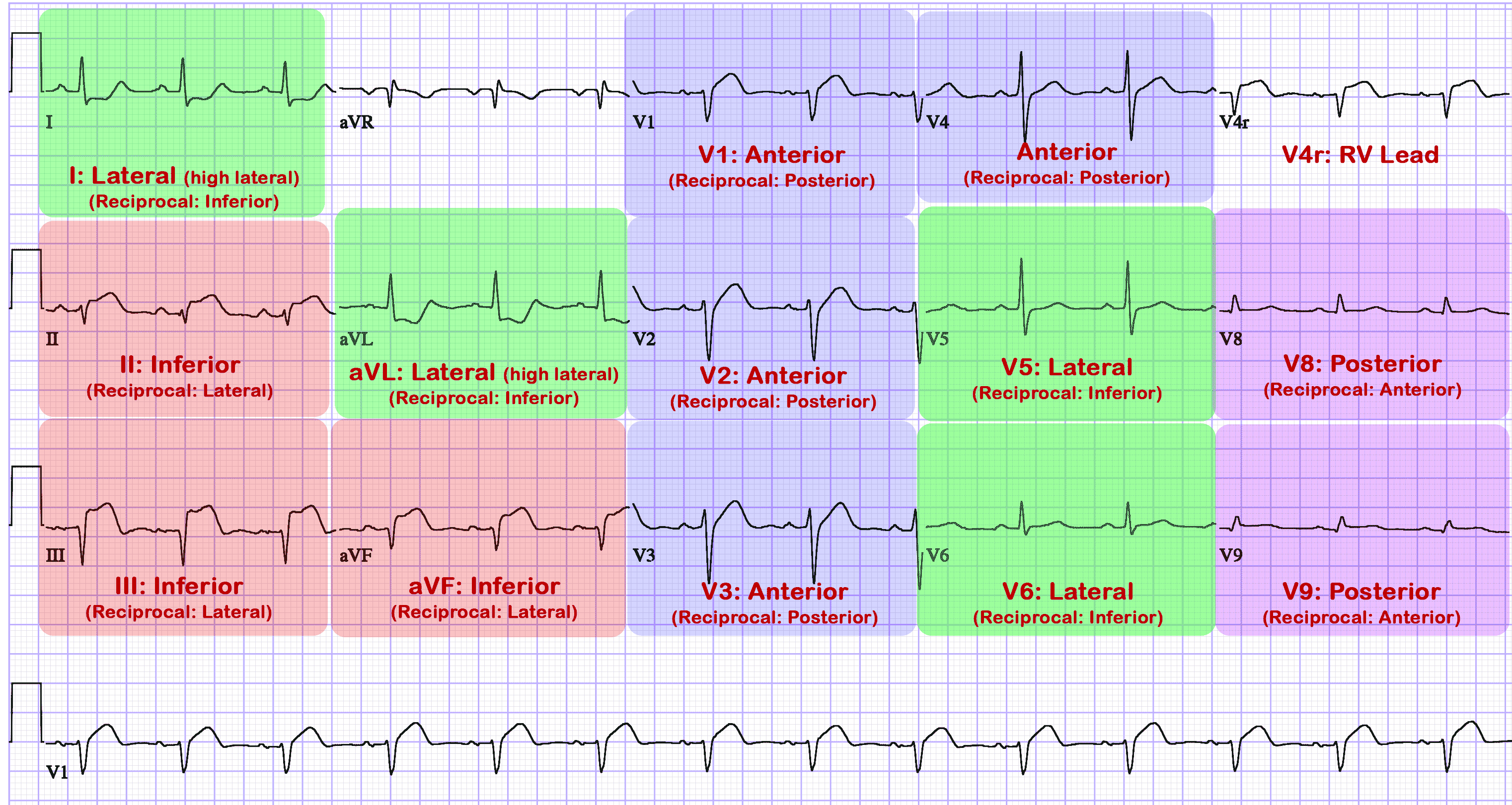
Stemi Leads Chart - For the ccrn one section of questions relies on your. This type of stemi usually occurs when a blockage occurs in the left anterior descending (lad) artery, the largest artery which provides blood flow. ≥0,5 mm, except from men patients with stemi the ecg leads displaying st segment elevations actually reflect the ischemic area.</strong> see more St elevation ≥1.5 mm in v2 or v3, or ≥1 mm in any. Look for a reciprocal stemi! ≥ 2 mm st segment elevation in precordial leads. Web home ecg library. Web learn how to diagnose and manage stemi, a true cardiac emergency, using ecg criteria, lead orientation, and sgarbossa criteria. The acceptable degree of st elevation in v2 and v3 changes based on age and gender. This badge card is a 12 lead stemi reference tool. St elevation ≥1.5 mm in v2 or v3, or ≥1 mm in any. Web this topic last updated: Web learn how to diagnose and manage stemi, a true cardiac emergency, using ecg criteria, lead orientation, and sgarbossa criteria. The acceptable degree of st elevation in v2 and v3 changes based on age and gender. Findings present in at least 2. The acceptable degree of st elevation in v2 and v3 changes based on age and gender. Look for a reciprocal stemi! Web home ecg library. ≥ 2 mm st segment elevation in precordial leads. St elevation ≥1.5 mm in v2 or v3, or ≥1 mm in any. Web apr 29, 2019. An easy way to memorize stemi locations and leads. The acceptable degree of st elevation in v2 and v3 changes based on age and gender. Follow the links above to find out more about the. For the ccrn one section of questions relies on your. Find out how to rule out. This badge card is a 12 lead stemi reference tool. Look for a reciprocal stemi! Web learn how to diagnose and manage stemi, a true cardiac emergency, using ecg criteria, lead orientation, and sgarbossa criteria. An easy way to memorize stemi locations and leads. ≥ 2 mm st segment elevation in precordial leads. •diagnostic elevation (in absence of lvh and lbbb) defined as: St elevation ≥1.5 mm in v2 or v3, or ≥1 mm in any. St elevation ≥2 mm in v2 or v3, or ≥1 mm in any other leads. Web st depression from subendocardial ischemia does not localise. An easy way to memorize stemi locations and leads. Web st depression from subendocardial ischemia does not localise. Ecg anatomy correlation mi localization. St elevation ≥1.5 mm in v2 or v3, or ≥1 mm in any. View a larger version of this infographic. For the ccrn one section of questions relies on your. Findings present in at least 2. ≥ 2 mm st segment elevation in precordial leads. It shows the standard 12 lead layout and the associated adjacent leads relative to the location in the heart. Web st depression from subendocardial ischemia does not localise. Web home ecg library. Ecg anatomy correlation mi localization. Web st depression from subendocardial ischemia does not localise. ≥0,5 mm, except from men patients with stemi the ecg leads displaying st segment elevations actually reflect the ischemic area.</strong> see more The acceptable degree of st elevation in v2 and v3 changes based on age and gender. View a larger version of this infographic. Men & women v4r and v3r: For the ccrn one section of questions relies on your. An easy way to memorize stemi locations and leads. This type of stemi usually occurs when a blockage occurs in the left anterior descending (lad) artery, the largest artery which provides blood flow. An easy way to memorize stemi locations and leads. •diagnostic elevation (in absence of lvh and lbbb) defined as: Web st depression from subendocardial ischemia does not localise. Men & women v4r and v3r: View a larger version of this infographic. ≥0,5 mm, except from men patients with stemi the ecg leads displaying st segment elevations actually reflect the ischemic area.</strong> see more •diagnostic elevation (in absence of lvh and lbbb) defined as: This badge card is a 12 lead stemi reference tool. Web home ecg library. View a larger version of this infographic. Web 1 mm of st elevation in any two contiguous leads except v2 and v3. Web apr 29, 2019. Findings present in at least 2. St elevation ≥2 mm in v2 or v3, or ≥1 mm in any other leads. Men & women v4r and v3r: St elevation ≥1.5 mm in v2 or v3, or ≥1 mm in any. Look for a reciprocal stemi! Find out how to rule out. Web learn how to diagnose and manage stemi, a true cardiac emergency, using ecg criteria, lead orientation, and sgarbossa criteria. The acceptable degree of st elevation in v2 and v3 changes based on age and gender. Follow the links above to find out more about the.
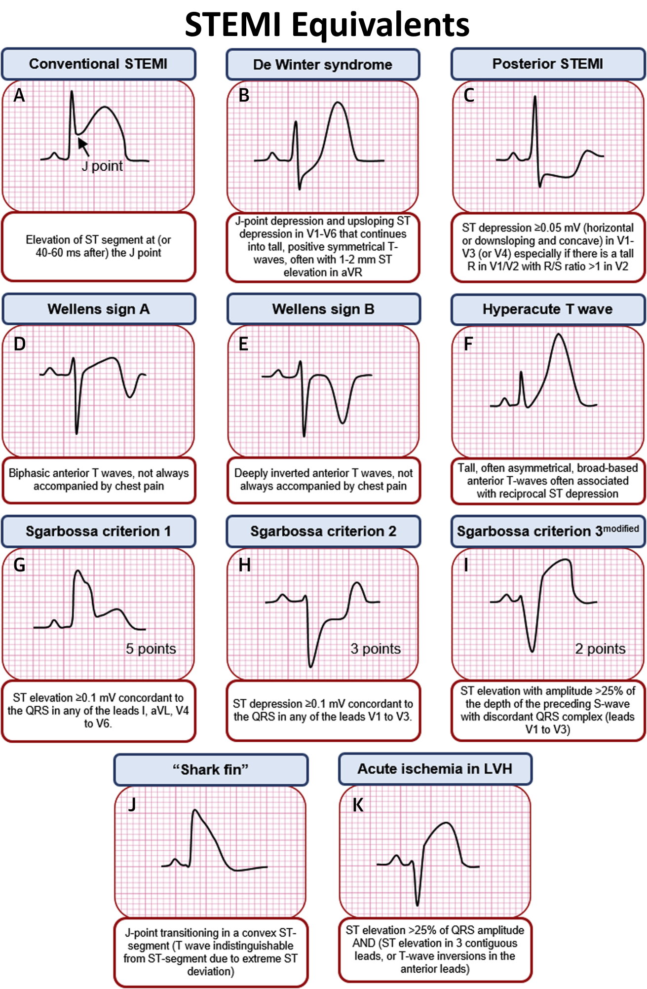
STEMI Equivalents on ECG • Conventional STEMI Elevation GrepMed

12 Lead Ekg Stemi Chart

12 Lead Stemi Chart

12 Lead STEMI Chart

ST Elevation MI (STEMI) Cardio Guide

Understanding 12 Lead Part2 LATERAL STEMI YouTube

STEMI & NSTEMI A Nurse's Comprehensive Guide Health And Willness

Pin on Nursing

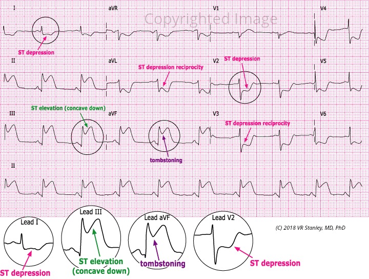
Acute Inferior STEMI Dr. Stanley's Dr. Stanley's

STEMI 12leads Nursing mnemonics, Emergency nursing, Nurse
For The Ccrn One Section Of Questions Relies On Your.
Web This Topic Last Updated:
An Easy Way To Memorize Stemi Locations And Leads.
≥1 Mm (0.1 Mv) St Segment Elevation In Limb Leads.
Related Post: