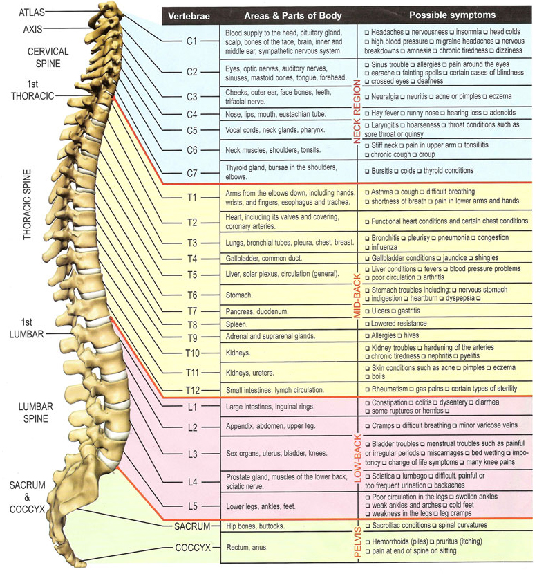
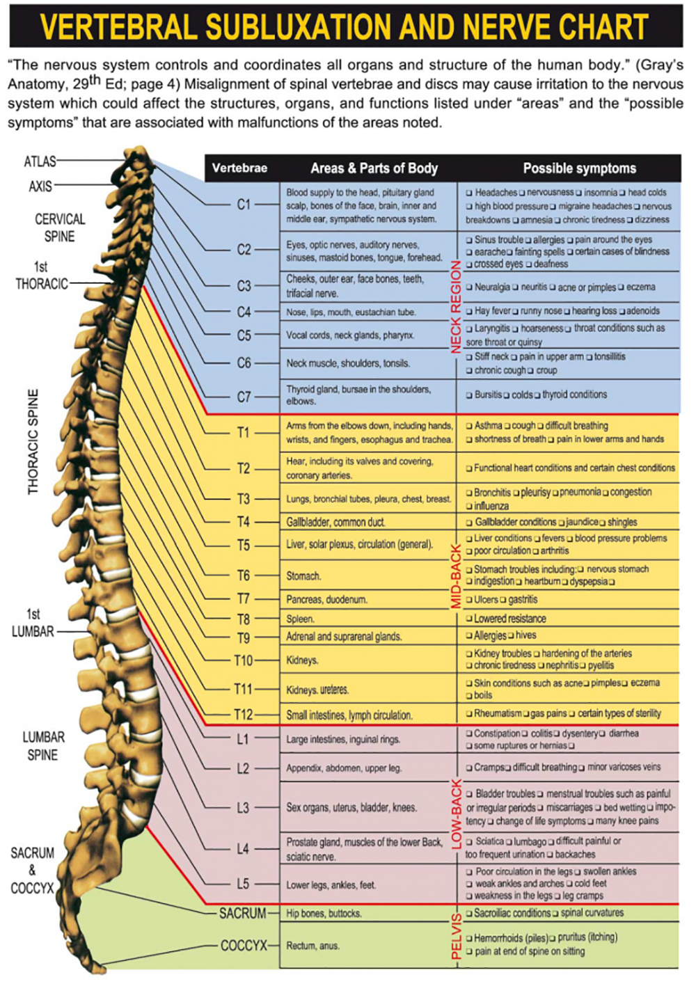
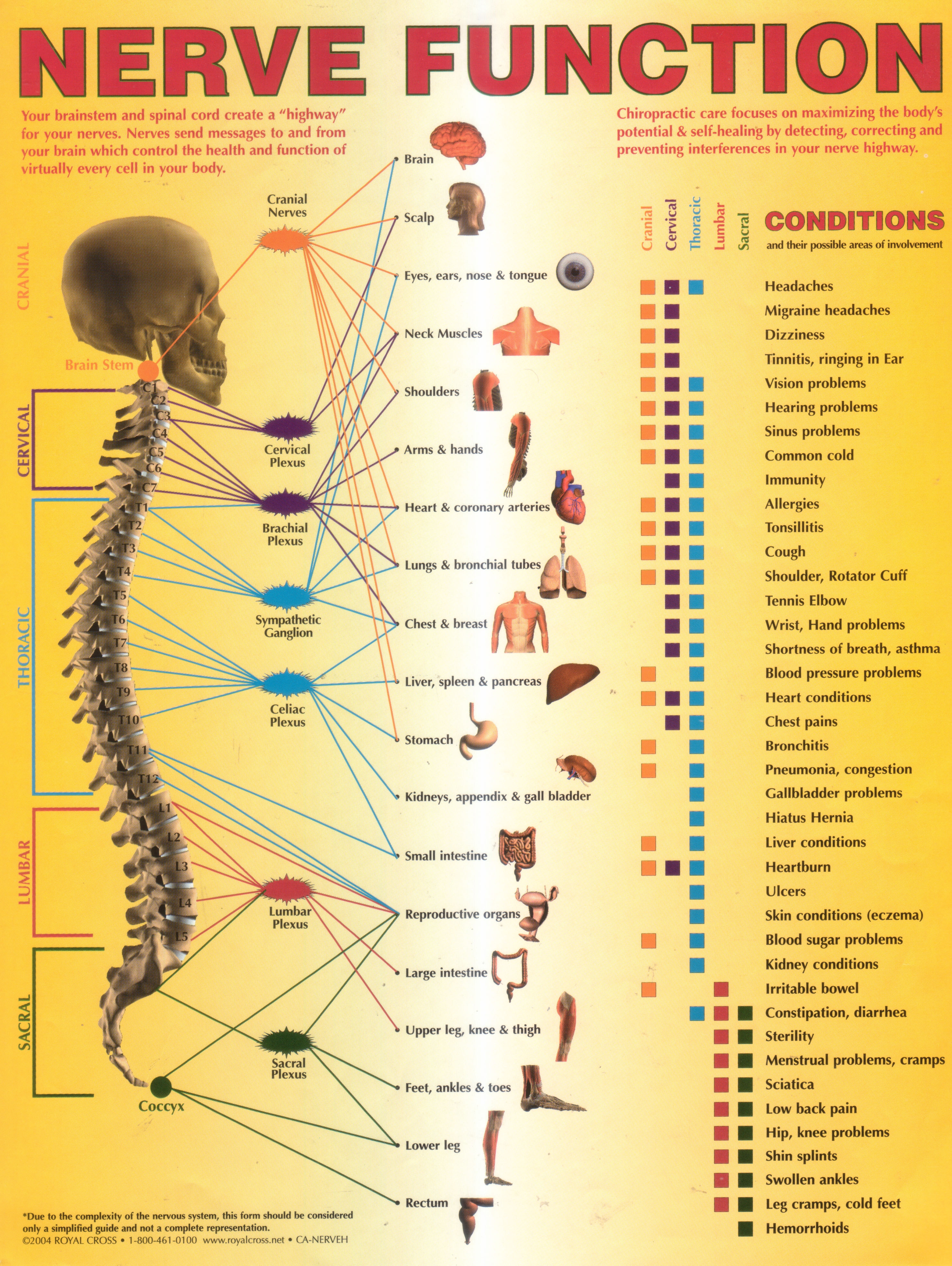
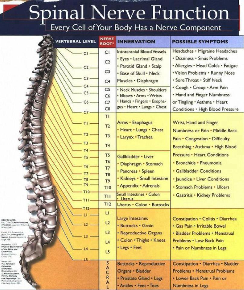
Spinal Nerve Chart
Spinal Nerve Chart - For the most part, the spinal nerves exit the vertebral canal through the intervertebral foramen below their corresponding vertebra. Web there are seven cervical vertebrae at the top, followed by 11 thoracic vertebrae, five lumbar vertebrae at the lower back, and five fused vertebrae at the bottom to create the sacrum. Spinal cord and spinal nerves. A dermatome is an area of skin supplied by a single spinal nerve. Web there are 5 pairs of lumbar spinal nerves that progressively increase in size from l1 to l5. This root has a swelling called the dorsal root ganglion, which contains the cell bodies of sensory neurons. Eight cervical spinal nerve pairs, 12 thoracic pairs , five lumbar pairs, five sacral pairs, and one coccygeal pair. Each spinal nerve originates from two roots: Carries sensory (afferent) information to the spinal cord. Web the 30 dermatomes explained and located. Web the 30 dermatomes explained and located. This root has a swelling called the dorsal root ganglion, which contains the cell bodies of sensory neurons. This diagram indicates the formation of a typical spinal nerve from the dorsal and ventral roots. There are 31 pairs of spinal nerves that branch out from the spinal cord. Web there are 31 bilateral. This root has a swelling called the dorsal root ganglion, which contains the cell bodies of sensory neurons. Eight cervical spinal nerve pairs, 12 thoracic pairs , five lumbar pairs, five sacral pairs, and one coccygeal pair. Web there are 31 bilateral pairs of spinal nerves, named from the vertebra they correspond to. The spinal nerves emerge laterally from the. A dermatome is an area of skin supplied by a single spinal nerve. The spinal nerves emerge laterally from the vertebral column with 8 in the cervical region, 12 in the thoracic region, 5 in the lumbar region, 5 in the sacral region, and 1 in the coccygeal region. Web there are 5 pairs of lumbar spinal nerves that progressively. Web there are seven cervical vertebrae at the top, followed by 11 thoracic vertebrae, five lumbar vertebrae at the lower back, and five fused vertebrae at the bottom to create the sacrum. Spinal cord and spinal nerves. For the most part, the spinal nerves exit the vertebral canal through the intervertebral foramen below their corresponding vertebra. Web the 30 dermatomes. Spinal cord and spinal nerves. Each spinal nerve originates from two roots: A dermatome is an area of skin supplied by a single spinal nerve. Web there are 5 pairs of lumbar spinal nerves that progressively increase in size from l1 to l5. Web there are 31 bilateral pairs of spinal nerves, named from the vertebra they correspond to. Numbers indicate the types of nerve fibers: Web figure 12.4.1 12.4. Carries sensory (afferent) information to the spinal cord. Web there are 31 bilateral pairs of spinal nerves, named from the vertebra they correspond to. There are 31 pairs of spinal nerves that branch out from the spinal cord. Numbers indicate the types of nerve fibers: This root has a swelling called the dorsal root ganglion, which contains the cell bodies of sensory neurons. Web there are seven cervical vertebrae at the top, followed by 11 thoracic vertebrae, five lumbar vertebrae at the lower back, and five fused vertebrae at the bottom to create the sacrum. Web the 30. The spinal nerves emerge laterally from the vertebral column with 8 in the cervical region, 12 in the thoracic region, 5 in the lumbar region, 5 in the sacral region, and 1 in the coccygeal region. Web there are 31 bilateral pairs of spinal nerves, named from the vertebra they correspond to. This root has a swelling called the dorsal. Carries sensory (afferent) information to the spinal cord. This root has a swelling called the dorsal root ganglion, which contains the cell bodies of sensory neurons. Numbers indicate the types of nerve fibers: There are 31 pairs of spinal nerves that branch out from the spinal cord. Web the 30 dermatomes explained and located. For the most part, the spinal nerves exit the vertebral canal through the intervertebral foramen below their corresponding vertebra. Spinal cord and spinal nerves. Web there are seven cervical vertebrae at the top, followed by 11 thoracic vertebrae, five lumbar vertebrae at the lower back, and five fused vertebrae at the bottom to create the sacrum. This diagram indicates the. Numbers indicate the types of nerve fibers: A dermatome is an area of skin supplied by a single spinal nerve. Web there are 5 pairs of lumbar spinal nerves that progressively increase in size from l1 to l5. The spinal nerves emerge laterally from the vertebral column with 8 in the cervical region, 12 in the thoracic region, 5 in the lumbar region, 5 in the sacral region, and 1 in the coccygeal region. Carries sensory (afferent) information to the spinal cord. Web there are seven cervical vertebrae at the top, followed by 11 thoracic vertebrae, five lumbar vertebrae at the lower back, and five fused vertebrae at the bottom to create the sacrum. Eight cervical spinal nerve pairs, 12 thoracic pairs , five lumbar pairs, five sacral pairs, and one coccygeal pair. Spinal cord and spinal nerves. For the most part, the spinal nerves exit the vertebral canal through the intervertebral foramen below their corresponding vertebra. Web there are 31 bilateral pairs of spinal nerves, named from the vertebra they correspond to. This root has a swelling called the dorsal root ganglion, which contains the cell bodies of sensory neurons. Web figure 12.4.1 12.4. There are 31 pairs of spinal nerves that branch out from the spinal cord.
Why Choose Us Optimal Spine Chiropractic

Blog Best Rated Chiropractor in New York

Nerve Chart. Great chart showing how spinal nerve irritation affects

Spinal Nerves Spinal nerve, Spinal nerves anatomy, Basic anatomy and

Annual World Spine Day Campaign Nerve Function Chart

Spinal Nerve Chart medschool doctor medicalstudent Image Credits

Spinal Nerve Function Anatomical Chart Anatomy Models and Anatomical

the spiral nerve function is shown in this manual for students to learn

Human Anatomy and Physiology Spinal Nerve Function

Recipes and Tips To Fight M.S. Spinal Nerve Function
This Diagram Indicates The Formation Of A Typical Spinal Nerve From The Dorsal And Ventral Roots.
These Nerves Exit The Intervertebral Foramina Below The Corresponding Vertebra.
Each Spinal Nerve Originates From Two Roots:
Web The 30 Dermatomes Explained And Located.
Related Post: