Leg Muscle Anatomy Chart
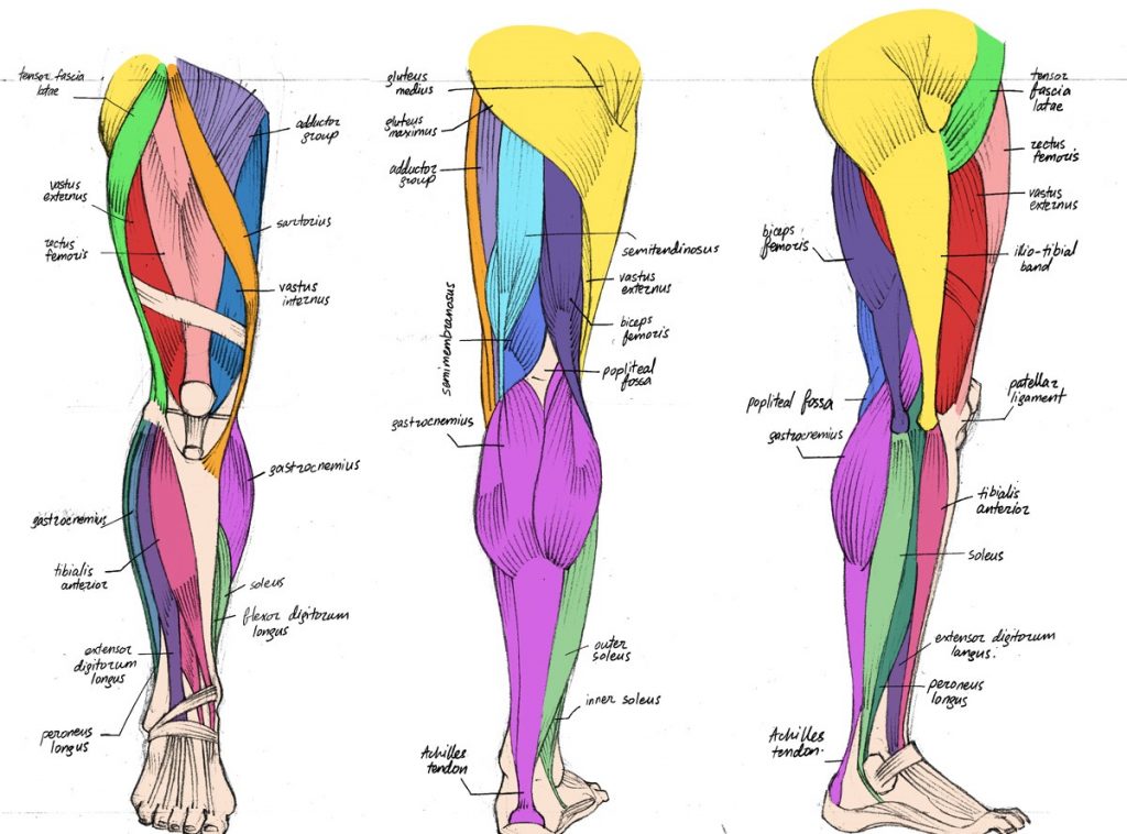
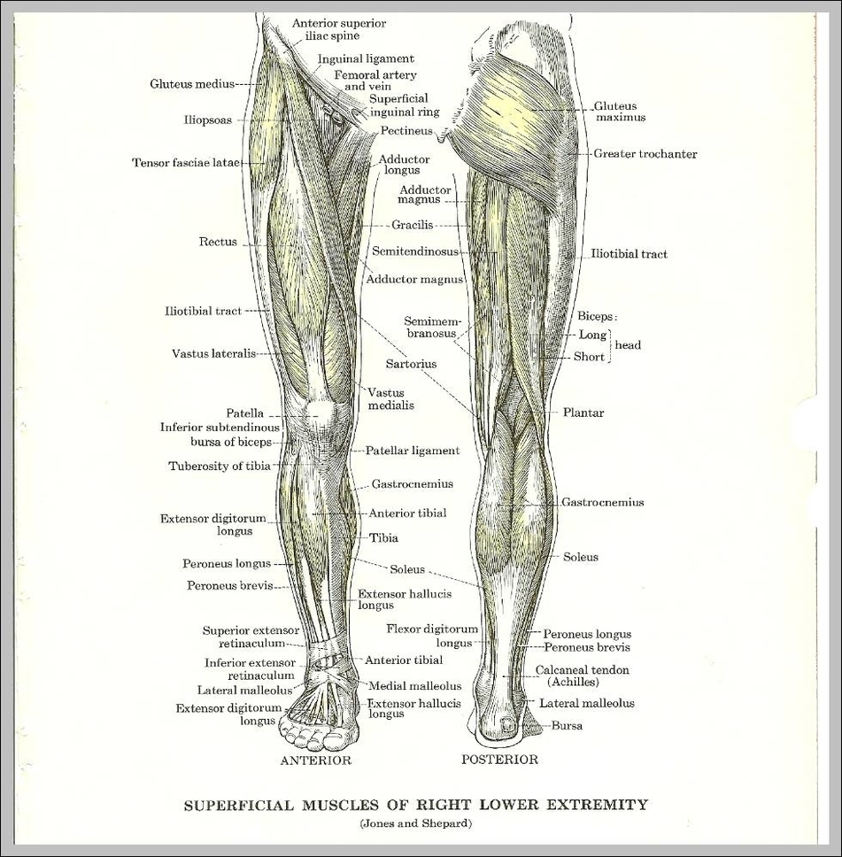
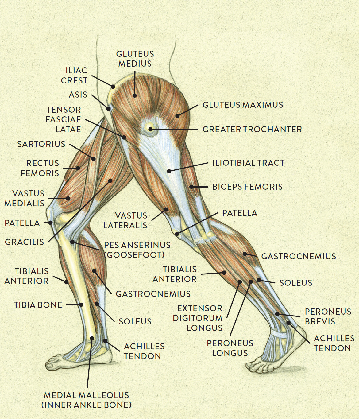
Leg Muscle Anatomy Chart - It is located toward the middle of the lower leg. The most visible and the largest glute muscle starts on your sacrum (the triangular bone at the base of the spine) and your lumbar fascia (connective tissue in your lower back) and attaches to your iliotibial tract, or your it band, and your outer thigh. Discover the muscle anatomy of every muscle group in the human body. Web when studying the muscles of the leg, they can be compartmentalized into four primary groups: Web three primary joints make up the structure of your lower half — your ankle, knee, and hip. This chart is beautifully illustrated and offers the most comprehensive look. Web muscles in the posterior compartment of the leg. Web your leg muscles work with your bones, tendons and ligaments to stabilize your body, support your weight and help you move. Each joint is encased by different muscles that perform unique functions and, most importantly, are. Last updated on october 27, 2019. The anterior compartment, the posterior compartment and the lateral compartment. The hamstrings are three muscles at the back of the thigh that affect hip and. The fibula and the tibia (shinbone). Anterior, lateral and posterior leg muscles; Web muscles of the leg. Web from the large, strong muscles of the buttocks and legs to the tiny, fine muscles of the feet and toes, these muscles can exert tremendous power while constantly making small adjustments for balance — whether the body is at rest or in motion. Inner hip & gluteal muscles; The most visible and the largest glute muscle starts on your. The most visible and the largest glute muscle starts on your sacrum (the triangular bone at the base of the spine) and your lumbar fascia (connective tissue in your lower back) and attaches to your iliotibial tract, or your it band, and your outer thigh. This is the largest quadriceps muscle. It is located toward the middle of the lower. Web these muscles are the adductor longus , adductor brevis , adductor magnus , gracilis, and the obturator externus. Web muscles of the leg. Web in the realm of anatomy, the ‘leg’ is strictly the region between the knee and the ankle joints rather than the entire lower extremity, as erroneously referred to in common language. Discover the muscle anatomy. The hip joint allows you to move and rotate your legs pelvic area in all directions. In this small section, we’ll briefly mention the main parts of the leg, namely the bones, muscles, and neurovasculature. It is available for free. The most visible and the largest glute muscle starts on your sacrum (the triangular bone at the base of the. You’ll learn about the muscles, bones, and other structures of each area of the leg. It is available for free. The fibula and the tibia (shinbone). Web master the leg muscle anatomy by exploring our videos, quizzes, labelled diagrams and articles. This muscle runs from front of your hip to the quadriceps tendon (the fibrous band above your kneecap), crosses. Learn about the three types of muscle as you use our 3d models to explore the anatomical structure and physiology of human muscles. Muscles help us move, and movement occurs in a three dimensional plane. Web true to their name, the quads consist of four major muscles of the leg: Web from the large, strong muscles of the buttocks and. Web these muscles are the adductor longus , adductor brevis , adductor magnus , gracilis, and the obturator externus. Web the muscles of the leg anatomy chart shows in every possible view the way that the muscles and other pieces of the leg work together in motion and flexibility. Web the leg anatomy is so complex, containing both the knee. In this page, we will take a look at all of the above as well as the anatomy of the knee. This is the largest quadriceps muscle. From a channel with a health professional licensed in germany. When it comes to learning about the anatomy and functions of muscles, 3d anatomical models surpass 2d learning experiences for one primary reason:. Find the best weight lifting exercises that target each muscle or groups of muscles. Web these muscles are the adductor longus , adductor brevis , adductor magnus , gracilis, and the obturator externus. The leg is defined anatomically as the portion of the lower limb from the knee joint to the ankle joint. Web it is composed of several structures:. Web your leg muscles work with your bones, tendons and ligaments to stabilize your body, support your weight and help you move. Web the majority of muscles in the leg are considered long muscles, in that they stretch great distances. The fibula and the tibia (shinbone). The hamstrings are three muscles at the back of the thigh that affect hip and. The anterior, lateral (fibular), superficial posterior, deep posterior compartments. As these muscles contract and relax, they move skeletal bones to create movement of the. Web these muscles are the adductor longus , adductor brevis , adductor magnus , gracilis, and the obturator externus. Web three primary joints make up the structure of your lower half — your ankle, knee, and hip. The tibia is stronger and more prominent than the fibula. It is available for free. Dorsal and plantar foot muscles; When it comes to learning about the anatomy and functions of muscles, 3d anatomical models surpass 2d learning experiences for one primary reason: And don't worry, we'll explain the naming of skeletal muscles, too. Web anatomy of the lower leg. Web we’ll break down the anatomy and function of the upper leg, knee, lower leg, ankle, and foot. Web true to their name, the quads consist of four major muscles of the leg:
Leg Muscle Anatomy Chart

muscular anatomy of thigh

BIO201Leg Muscles Leg muscles anatomy, Muscle anatomy, Anatomy

Muscles Of The Leg Laminated Anatomy Chart studiosixsound.co.za

anatomy of the legs

Leg Tendons Diagram
:background_color(FFFFFF):format(jpeg)/images/article/en/muscle-anatomy-charts/2mzZyy0wDF3gODSlIc8Okg_sample_page_1.png)
Muscles Diagrams Diagram Of Muscles And Anatomy Charts

Lower Leg Muscle Diagram Diagram Quizlet

Muscles of the Leg and Foot Classic Human Anatomy in Motion The

Muscles of lower leg Digman Fitness
Your Muscles In The Lower Leg Are Supported By Two Very Strong, Long Bones:
The Leg Is Defined Anatomically As The Portion Of The Lower Limb From The Knee Joint To The Ankle Joint.
In Total, There Are 13 Separate Muscles Across These Three Compartments.
The Most Visible And The Largest Glute Muscle Starts On Your Sacrum (The Triangular Bone At The Base Of The Spine) And Your Lumbar Fascia (Connective Tissue In Your Lower Back) And Attaches To Your Iliotibial Tract, Or Your It Band, And Your Outer Thigh.
Related Post: