Gram Positive And Negative Bacteria Chart
Gram Positive And Negative Bacteria Chart - Gram negative bacteria have cell walls with a thin layer of peptidoglycan. Actinomyces, bacillus, clostridium, corynebacterium, enterococcus, gardnerella, lactobacillus, listeria, mycoplasma, nocardia, staphylococcus, streptococcus, streptomyces ,etc. What does it mean if bacteria are gram negative? Would you like to start learning session with this topic items scheduled for future? They also cause different types of infections, and different types of antibiotics are effective against them. A gram stain test, which involves a chemical dye, stains the bacterium’s cell wall purple. They don’t retain crystal violet, so are stained red or pink. Web these are their key characteristics: Web most bacteria can be broadly classified as gram positive or gram negative. Differences in cell wall structure between these categories. Web you are done for today with this topic. Gram project is a medical education resource website containing diagrams, tables and flowcharts for all your quick referencing, revision and teaching needs. They also cause different types of infections, and different types of antibiotics are effective against them. In a gram stain test, bacteria are washed with a decolorizing solution after. They have thick cell walls. Differences in cell wall structure between these categories. Gram positive bacteria have cell walls composed of thick layers of peptidoglycan. Web gram positive bacteria types and classification. They also cause different types of infections, and different types of antibiotics are effective against them. In a gram stain test, bacteria are washed with a decolorizing solution after being dyed with crystal violet. Gram negative bacteria lack this thick coating. The difference between the two groups is believed to be due to a much larger peptidoglycan (cell wall) in gram positives. What does it mean if bacteria are gram negative? Gram positive cells stain purple. A gram stain test, which involves a chemical dye, stains the bacterium’s cell wall purple. Gram positive cells stain purple when subjected to a gram stain procedure. They also cause different types of infections, and different types of antibiotics are effective against them. Would you like to start learning session with this topic items scheduled for future? Differences in cell. Gram negative bacteria lack this thick coating. Would you like to start learning session with this topic items scheduled for future? Web you are done for today with this topic. Web gram positive bacteria: Gram project is a medical education resource website containing diagrams, tables and flowcharts for all your quick referencing, revision and teaching needs. In a gram stain test, bacteria are washed with a decolorizing solution after being dyed with crystal violet. Gram positive bacteria have a thick coating of peptidoglycan and stain purple with crystal violet. The bacterial cell wall of these organisms have thick peptidoglycan layers, which take up the purple/violet stain. Gram negative bacteria have cell walls with a thin layer. In a gram stain test, bacteria are washed with a decolorizing solution after being dyed with crystal violet. Web here is a look at the differences between gram positive and gram negative bacteria and why telling them apart is important. The difference between the two groups is believed to be due to a much larger peptidoglycan (cell wall) in gram. Web these are their key characteristics: Web you are done for today with this topic. They have thick cell walls. Differences in cell wall structure between these categories. Gram project is a medical education resource website containing diagrams, tables and flowcharts for all your quick referencing, revision and teaching needs. They don’t retain crystal violet, so are stained red or pink. Web you are done for today with this topic. They also cause different types of infections, and different types of antibiotics are effective against them. Gram positive bacteria have a thick coating of peptidoglycan and stain purple with crystal violet. Differences in cell wall structure between these categories. In a gram stain test, bacteria are washed with a decolorizing solution after being dyed with crystal violet. A gram stain test, which involves a chemical dye, stains the bacterium’s cell wall purple. Gram positive cells stain purple when subjected to a gram stain procedure. Gram positive bacteria have cell walls composed of thick layers of peptidoglycan. Gram positive bacteria. Web you are done for today with this topic. Web these are their key characteristics: Differences in cell wall structure between these categories. Gram positive cells stain purple when subjected to a gram stain procedure. In a gram stain test, bacteria are washed with a decolorizing solution after being dyed with crystal violet. Gram positive bacteria have a thick coating of peptidoglycan and stain purple with crystal violet. Web gram positive bacteria: What does it mean if bacteria are gram negative? Gram project is a medical education resource website containing diagrams, tables and flowcharts for all your quick referencing, revision and teaching needs. Gram negative bacteria have cell walls with a thin layer of peptidoglycan. Web most bacteria can be broadly classified as gram positive or gram negative. Would you like to start learning session with this topic items scheduled for future? The bacterial cell wall of these organisms have thick peptidoglycan layers, which take up the purple/violet stain. The difference between the two groups is believed to be due to a much larger peptidoglycan (cell wall) in gram positives. They also cause different types of infections, and different types of antibiotics are effective against them. Gram project is a medical education resource website containing diagrams, tables and flowcharts for all your quick referencing, revision and teaching needs.
Gram Positive Vs Gram Negative Antibiotics
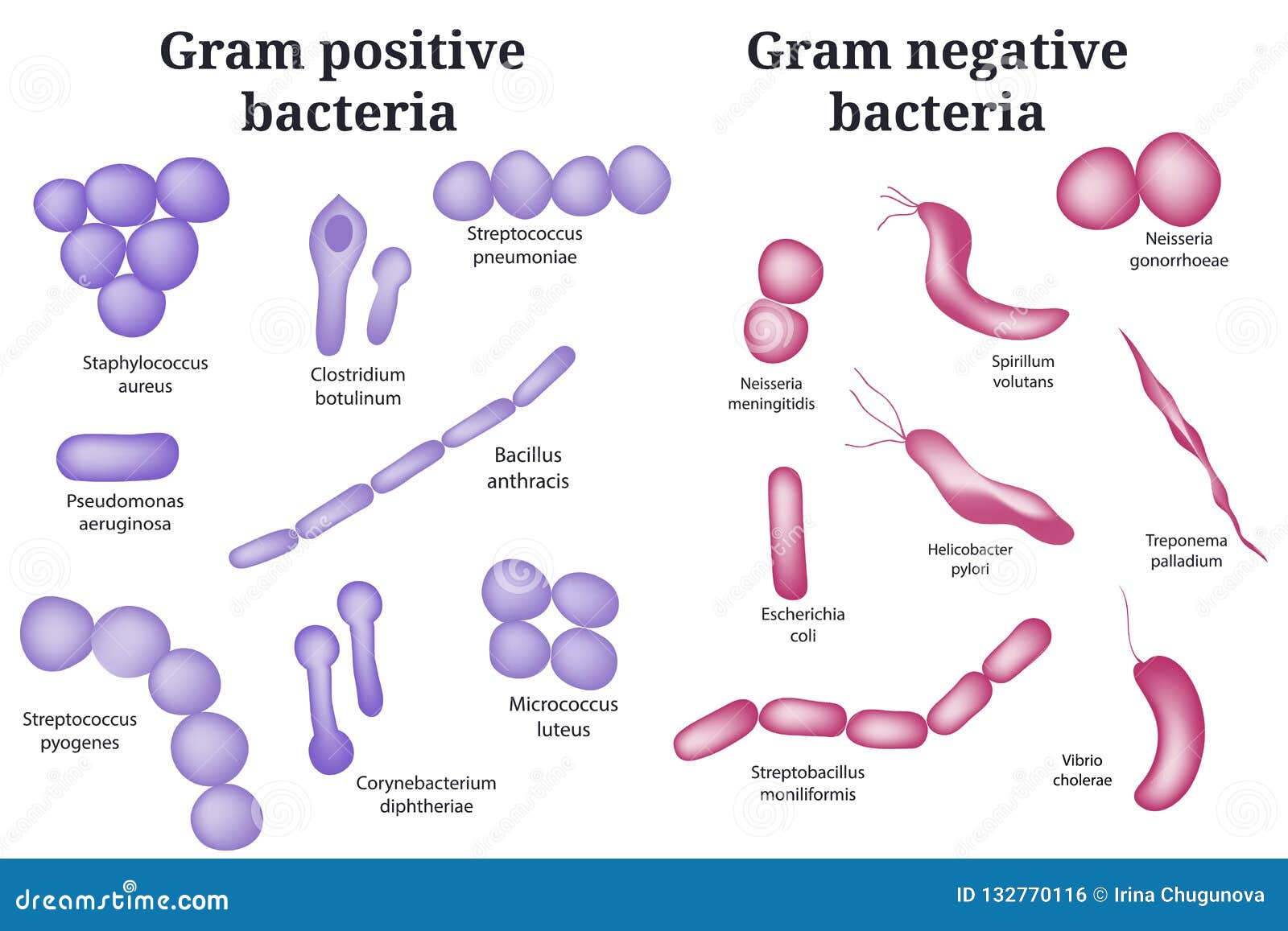
Gram Positive and Gram Negative Bacteria. Stock Vector Illustration
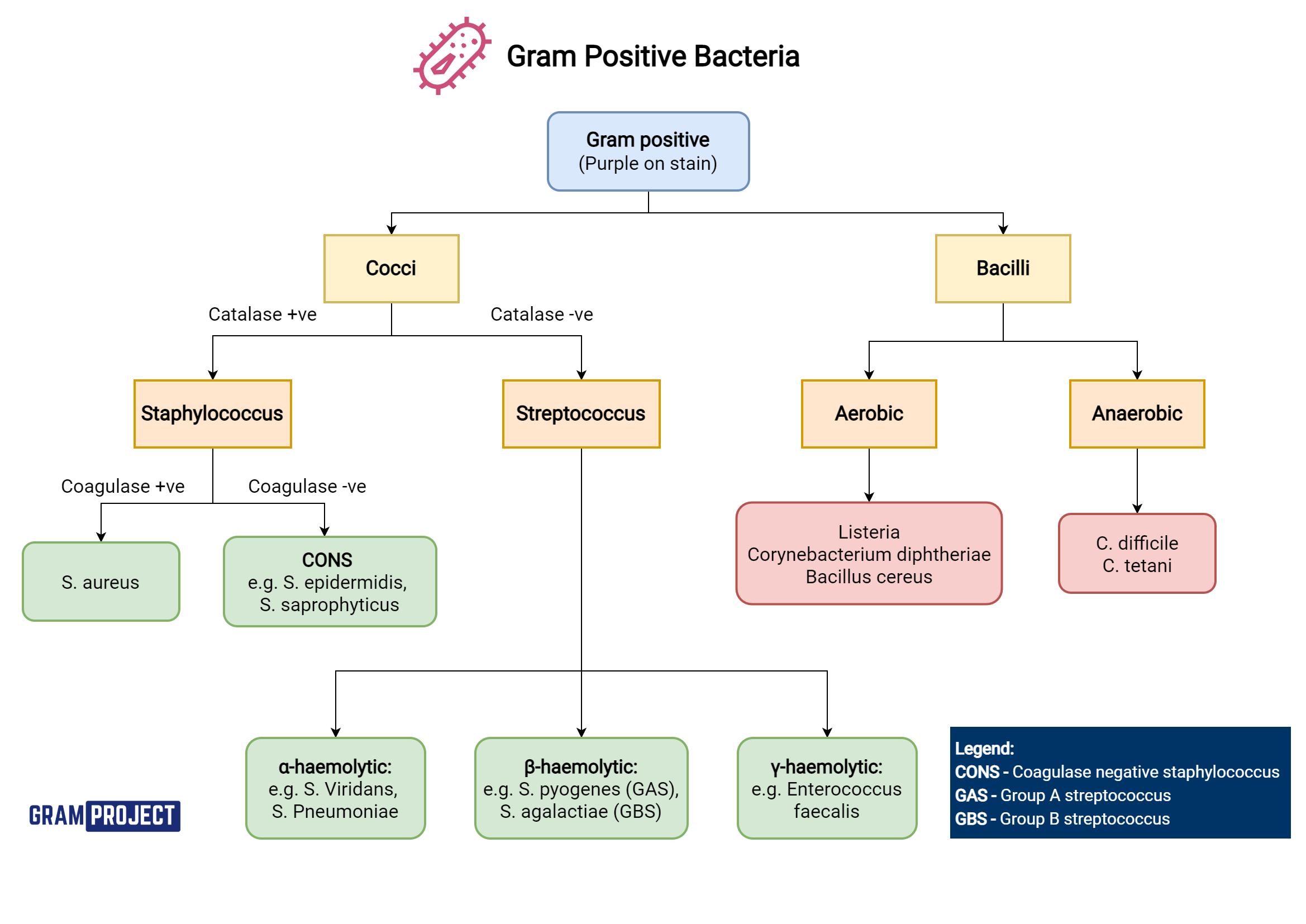
Gram Positive Organisms Chart

Gram Positive Vs Gram Negative Bacteria Chart
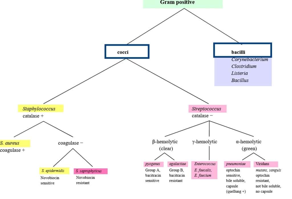
List Of Gram Positive And Negative Bacteria

ClASSIFICATION OF BACTERIA ON BASIS OF GRAM STAIN Google search
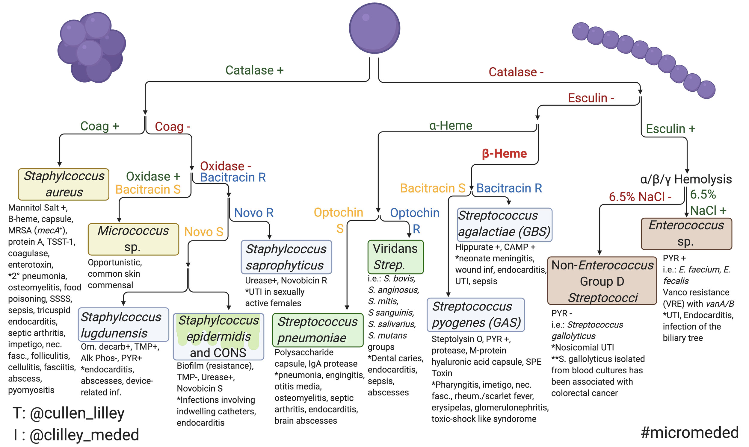
Gram Negative Bacteria Chart

Classification of Gram Positive&Negative bacteria by Organs. 7B
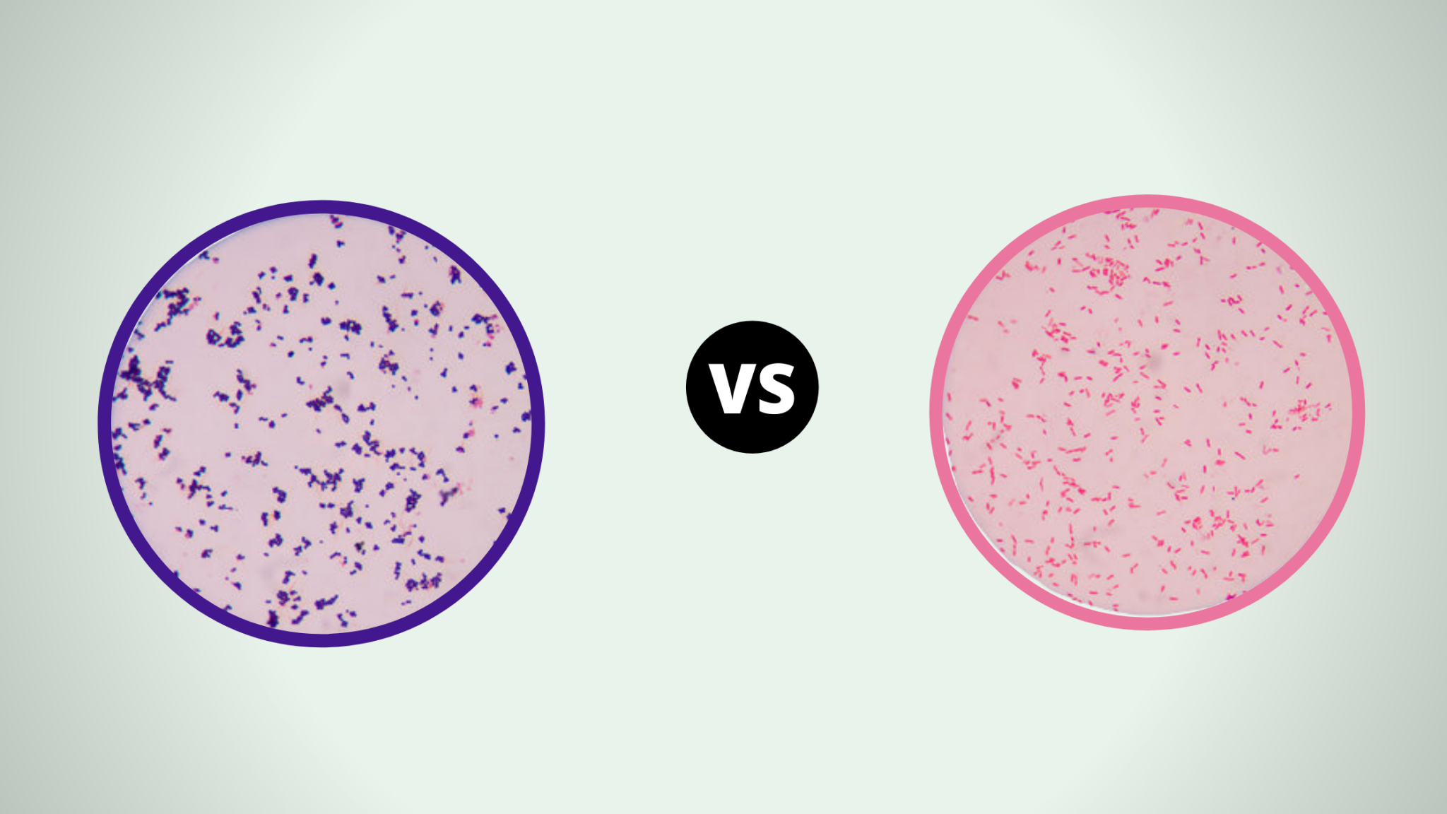
Gram Positive Vs Gram Negative Bacteria Chart

bacteria Medical technology, Medical laboratory science, Microbiology
A Gram Stain Test, Which Involves A Chemical Dye, Stains The Bacterium’s Cell Wall Purple.
Web May 29, 2024.
Actinomyces, Bacillus, Clostridium, Corynebacterium, Enterococcus, Gardnerella, Lactobacillus, Listeria, Mycoplasma, Nocardia, Staphylococcus, Streptococcus, Streptomyces ,Etc.
They Have Thick Cell Walls.
Related Post: