Foot Anatomy Chart
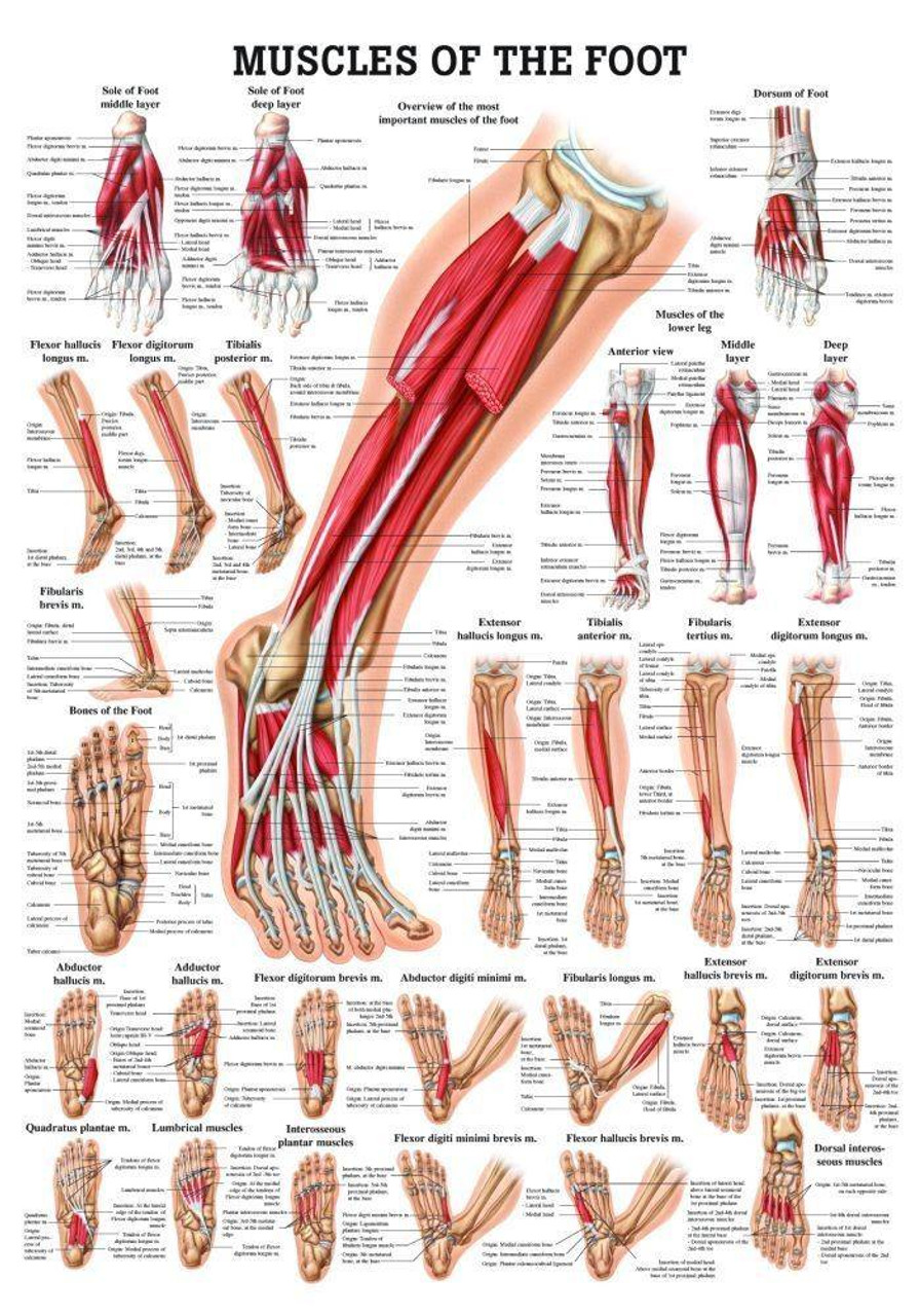
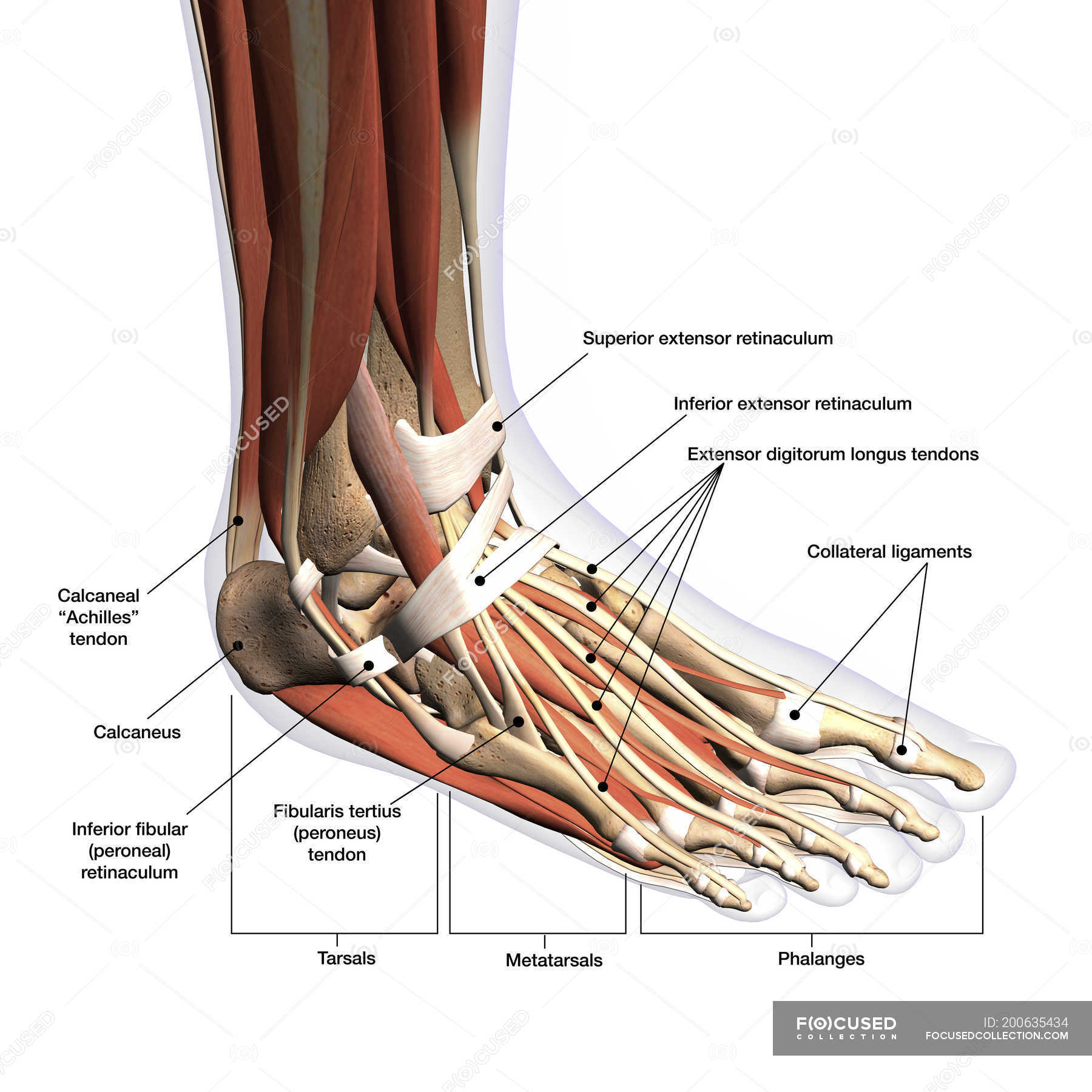
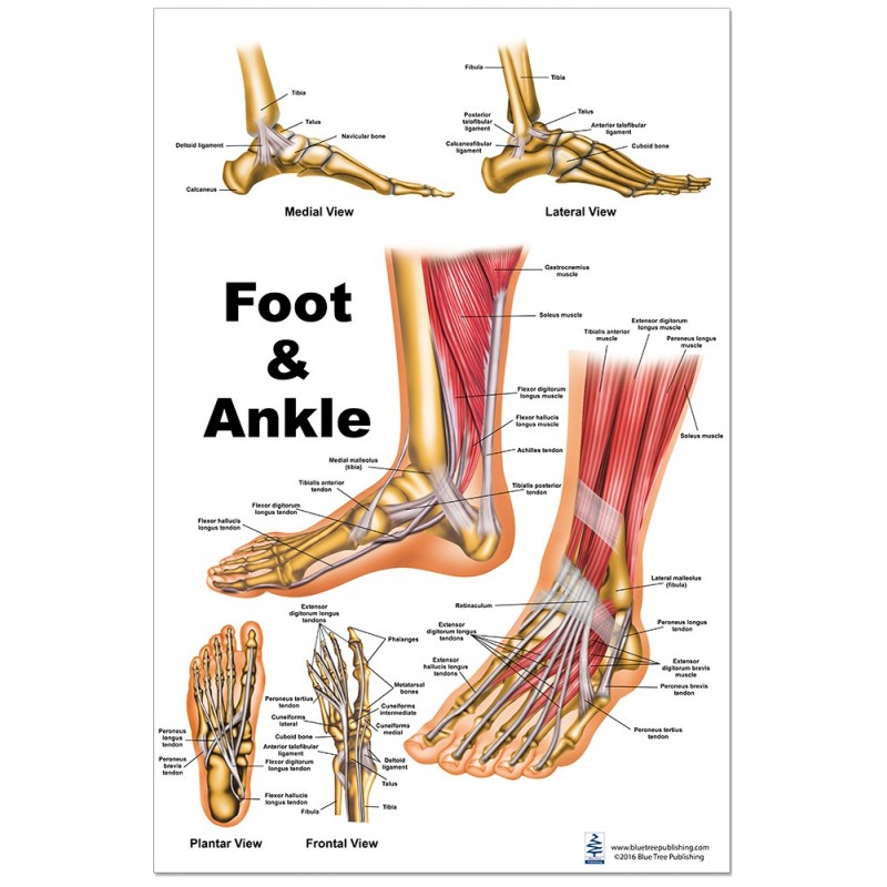
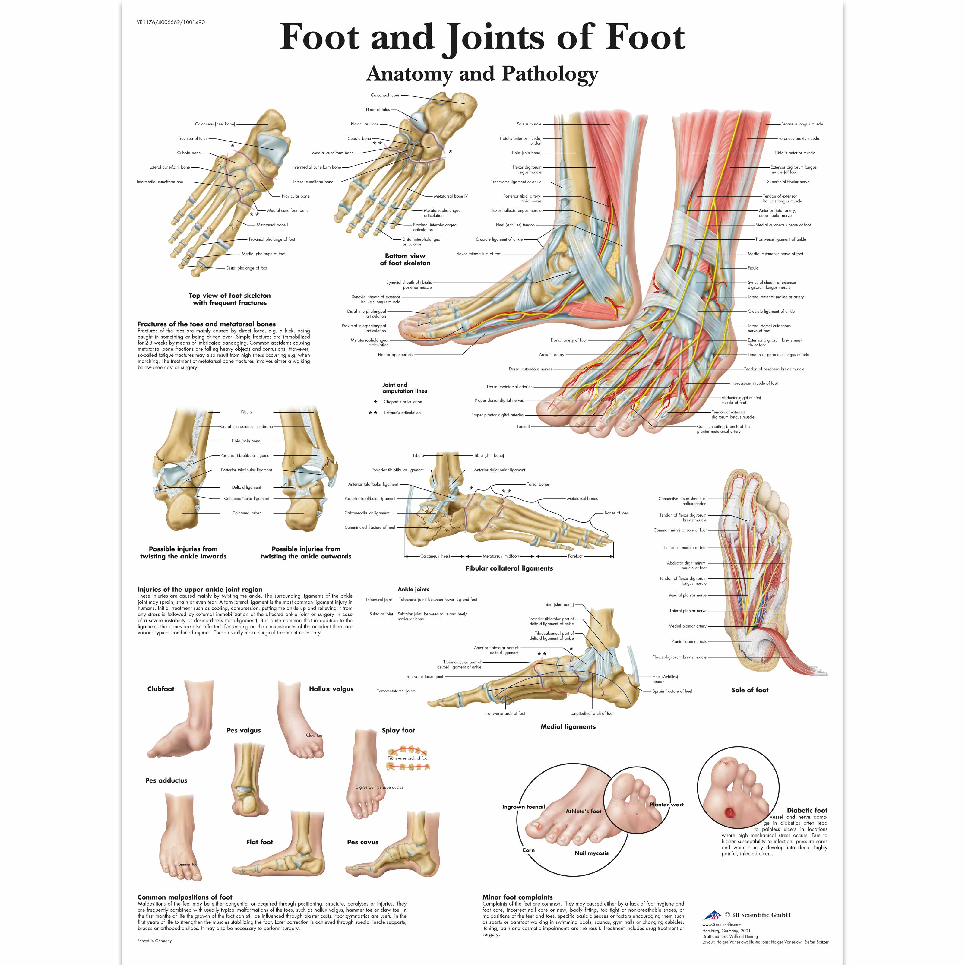
Foot Anatomy Chart - The midfoot, containing the navicular, cuboid, and cuneiforms; Web this article will outline some of the main anatomical features of the foot. These bones give structure to the foot and allow for all foot movements like flexing the toes and ankle, walking, and running. These bones are divided into three main regions: Web the anatomy of feet: The ankle or tibiotalar joint constitutes the junction of the lower leg and foot. Web in humans, the foot is one of the most complex structures in the body. The dorsal layer of foot muscles. Like the fingers, the toes have flexor and extensor muscles that power. There are ten intrinsic muscles located in the plantar aspect (sole) of the foot. Upper ankle joint (tibiotarsal), talocalcaneonavicular, and subtalar joints. Web the anatomy of feet: It will also look at some of the common conditions that affect the foot and their possible treatment options. The last two together are called the lower ankle joint. Web there are a variety of anatomical structures that make up the anatomy of the foot and ankle. How to ease foot pain at home. Like the fingers, the toes have flexor and extensor muscles that power. Major (2nd most important) medial arch support. [1] [2] the medial compartment contains the abductor hallucis, flexor hallicus brevis, and. Upper ankle joint (tibiotarsal), talocalcaneonavicular, and subtalar joints. These bones give structure to the foot and allow for all foot movements like flexing the toes and ankle, walking, and running. The dorsal layer of foot muscles. Bones of the foot and ankle. Web the anatomy of feet: These will be reviewed in the sections of this chapter. These will be reviewed in the sections of this chapter. The anatomic structures below the ankle joint comprise the foot, which includes: Web the many bones, ligaments, and tendons of the foot help you move, but they can also be injured and limit your mobility. This article will give an overview of foot anatomy and foot problems that come from. Web there are a variety of anatomical structures that make up the anatomy of the foot and ankle (figure 1) including bones, joints, ligaments, muscles, tendons, and nerves. These bones are divided into three main regions: The last two together are called the lower ankle joint. Web now that you’ve measured your foot size, compare the measurements against a nike. Web the foot muscles are divided into nine compartments encompassed by fascia, although past studies on the exact divisions of the foot have varied considerably. The last two together are called the lower ankle joint. Like the fingers, the toes have flexor and extensor muscles that power. Upper ankle joint (tibiotarsal), talocalcaneonavicular, and subtalar joints. This complex network of structures. The midfoot, containing the navicular, cuboid, and cuneiforms; The ankle joint, also known as the talocrural joint, allows dorsiflexion and plantar flexion of the foot. The ankle or tibiotalar joint constitutes the junction of the lower leg and foot. Web it consists of 28 bones, which can be divided functionally into three groups, referred to as the tarsus, metatarsus and. The osseous components of the ankle joint include the distal tibia, distal fibula, and talus. The foot is not only complicated in terms of the number and structure of bones, but also in terms of its joints. Upper ankle joint (tibiotarsal), talocalcaneonavicular, and subtalar joints. [1] [2] the medial compartment contains the abductor hallucis, flexor hallicus brevis, and. The last. [3] [4] this article discusses the key anatomical structures of the foot. There are ten intrinsic muscles located in the plantar aspect (sole) of the foot. A clinician's ability to understand the anatomical structures of the foot is crucial for assessment and treatment, especially for clinicians working with clients with musculoskeletal conditions. The hindfoot, midfoot, and forefoot. Bones of the. The hindfoot, midfoot, and forefoot. Web now that you’ve measured your foot size, compare the measurements against a nike size chart to find the best shoe size for you. [1] [2] the medial compartment contains the abductor hallucis, flexor hallicus brevis, and. This article will give an overview of foot anatomy and foot problems that come from overuse, injury, and. Upper ankle joint (tibiotarsal), talocalcaneonavicular, and subtalar joints. Major (2nd most important) medial arch support. These bones are divided into three main regions: Learn more about foot bones and foot anatomy here. This article will give an overview of foot anatomy and foot problems that come from overuse, injury, and normal wear and tear of the foot. [1] [2] the medial compartment contains the abductor hallucis, flexor hallicus brevis, and. Web this article will outline some of the main anatomical features of the foot. Ebraheim’s educational animated video describes anatomical structures of the foot and ankle, the bony anatomy, the joints, ligaments, and the compartments, in a simple and easy way. While other shoe brands may use similar size scales, it’s best to check out a shoe brand’s size charts before purchasing a new pair of shoes from them, as each brand’s exact approach to size and fit may vary. Web the anatomy of feet: Web foot and ankle anatomy consists of 33 bones, 26 joints and over a hundred muscles, ligaments and tendons. A clinician's ability to understand the anatomical structures of the foot is crucial for assessment and treatment, especially for clinicians working with clients with musculoskeletal conditions. The last two together are called the lower ankle joint. Base of the 5th metatarsal (lateral band), plantar plate and bases of the five proximal phalanges. The midfoot, containing the navicular, cuboid, and cuneiforms; The ankle or tibiotalar joint constitutes the junction of the lower leg and foot.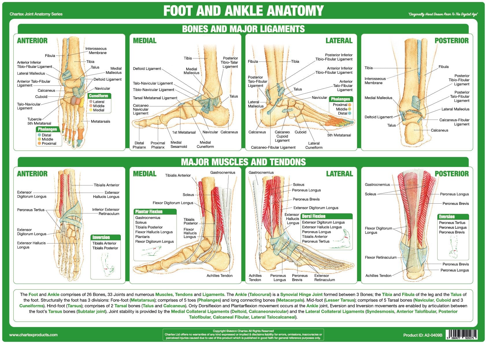
hand bone anatomy joints
Foot Ankle Anatomy Chart Poster Laminated ubicaciondepersonas.cdmx.gob.mx

Foot Anatomy 101 A Quick Lesson From a New Hampshire Podiatrist Nagy
Anatomy of human foot with labels on white background — ankle, leg

Foot & Ankle Anatomy Chart Feet Poster Anatomical Chart

This chart shows foot and ankle bone and ligament anatomy, normal
Foot Muscles Anatomy lupon.gov.ph

Understanding the Foot and Ankle 1004 Anatomical Parts & Charts
Anatomical Charts and Posters Anatomy Charts Foot and Ankle

hand bone and tendon chart Artist The Dorsal Foot How Do I Love
It Is Made Up Of Three Joints:
Web The Foot Is Divided Into Three Parts:
The Foot Is Not Only Complicated In Terms Of The Number And Structure Of Bones, But Also In Terms Of Its Joints.
[3] [4] This Article Discusses The Key Anatomical Structures Of The Foot.
Related Post: