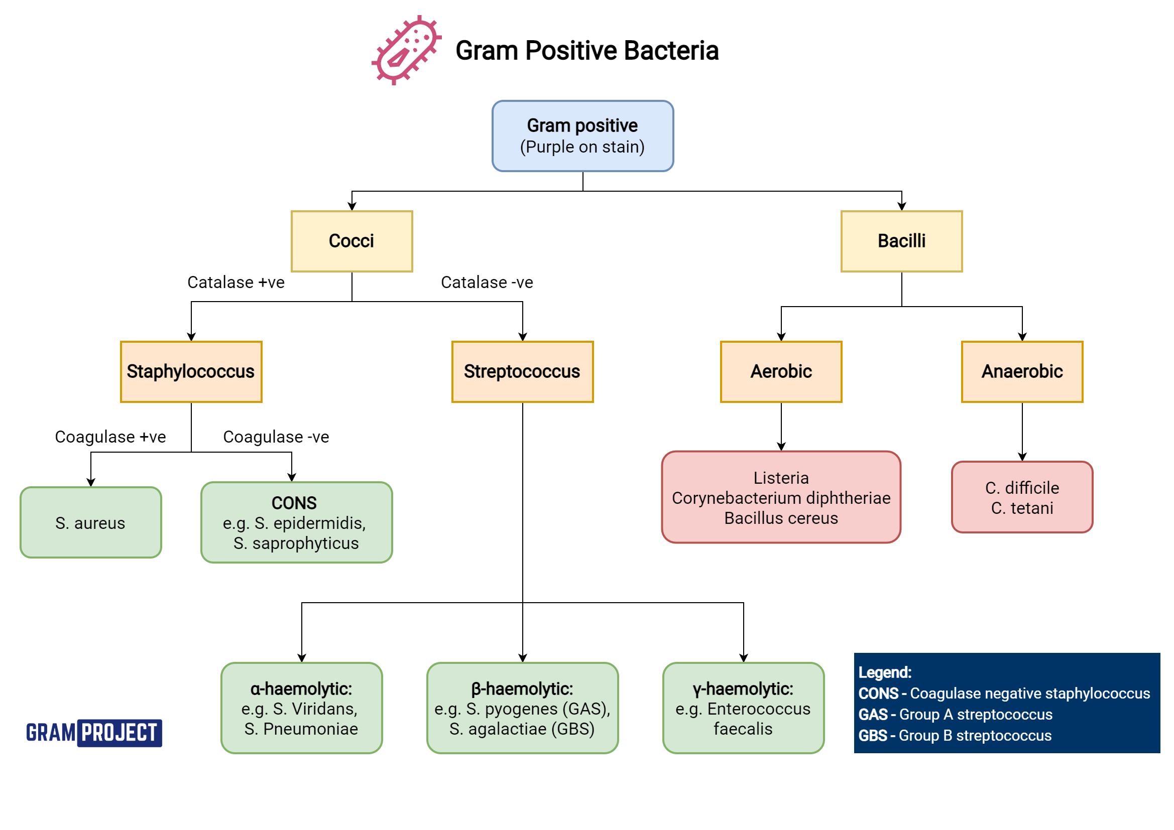
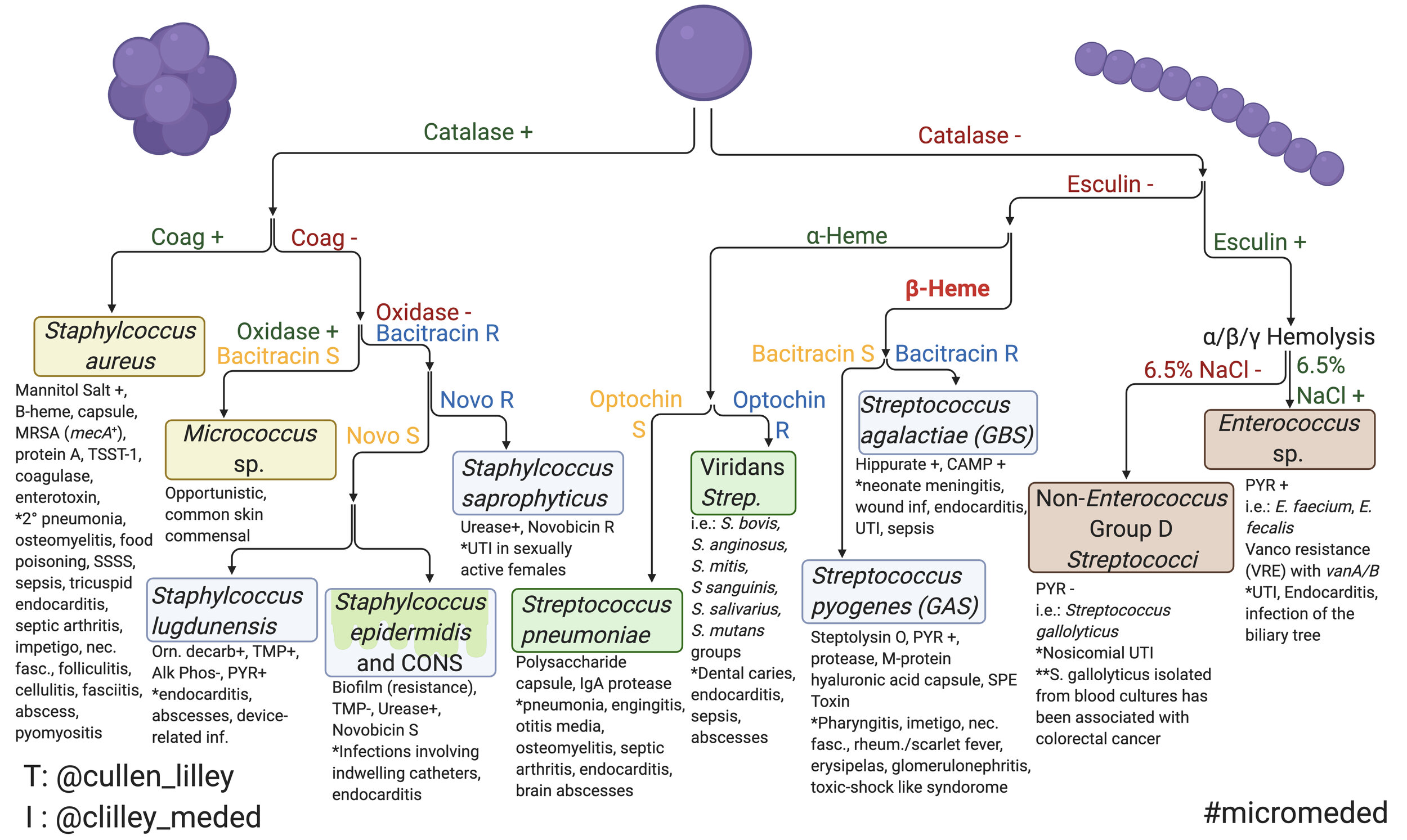
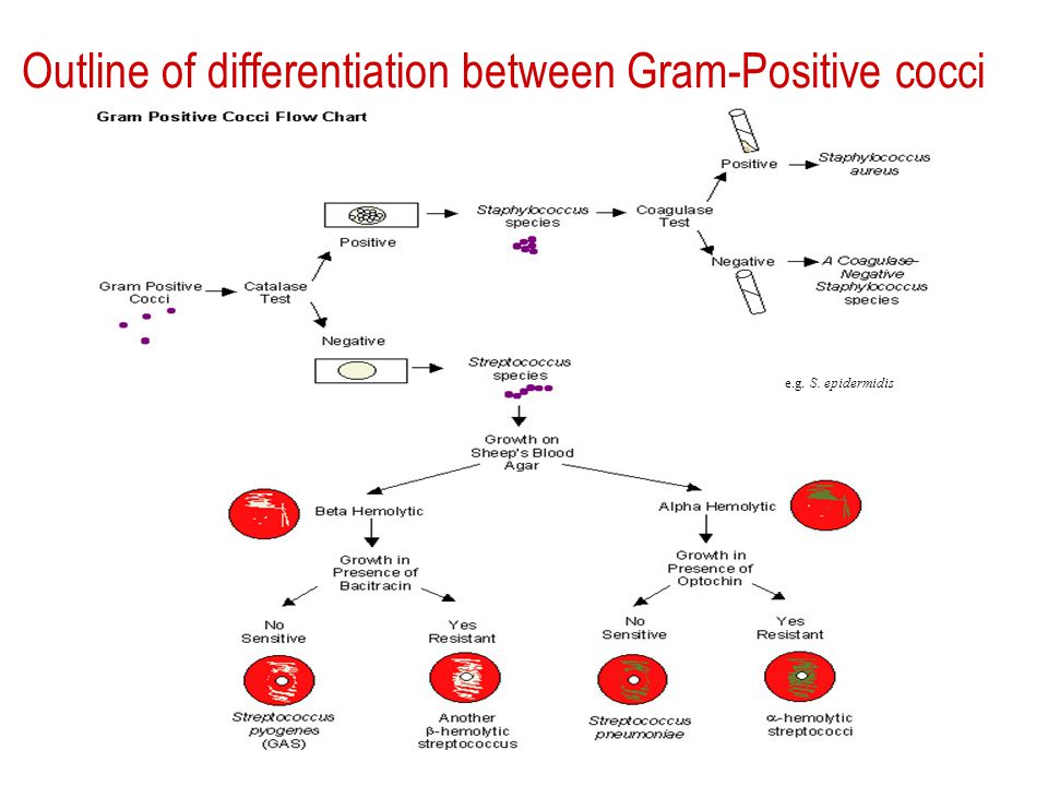
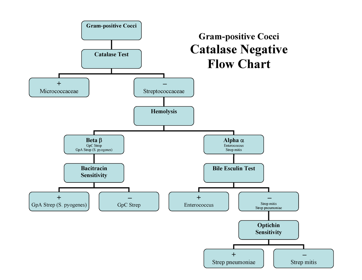
Flow Chart Of Gram Positive Cocci
Flow Chart Of Gram Positive Cocci - Web guide to interpretation of positive blood cultures i arrange the potential gram stain results that one can be called w/ as follows: Web this paper reviewed core concepts of interpreting bacterial culture results, including timing of cultures, common culture sites, potential for contamination, interpreting the gram stain, role of rapid diagnostic tests, conventional antibiotic susceptibility testing, and automated testing. 1 is a flow chart developed to show the key distinguishing tests. Web gram positive cocci obligate anaerobic peptostreptococcus spp., peptinophilus spp., parvimonas spp., anaerococcus spp., atopobium spp., f. Web aerobic gram positive cocci flowchart. Spheres (cocci), rods (bacilli), and spirals or helixes (spirochetes). Two of the most important human pathogens, staphylococcus aureus and streptococcus pyogenes , are described in this chapter. Coagulase negative staphylococci in multiple blood cultures. Give an example of a bacterium of high g+c and low g+c group commonly associated with each category. Mechanisms of antibiotic resistance in enterococci. Other tests not shown in the flow chart but included in table 2 can be used for. Causative agent for endocarditis and dental abscesses. Web aerobic gram positive cocci flowchart. Oxygen requirements, morph., growth requirements (45°c and supplements), read genera descriptions. Web back gram positive cocci. Web this paper reviewed core concepts of interpreting bacterial culture results, including timing of cultures, common culture sites, potential for contamination, interpreting the gram stain, role of rapid diagnostic tests, conventional antibiotic susceptibility testing, and automated testing. Web gram positive cocci obligate anaerobic peptostreptococcus spp., peptinophilus spp., parvimonas spp., anaerococcus spp., atopobium spp., f. Coagulase negative staphylococci in multiple blood. Causative agent for endocarditis and dental abscesses. Give an example of a bacterium of high g+c and low g+c group commonly associated with each category. Web the six flow charts we’ll be discussing are: Bacteria are identified in laboratories by various methods, including microscopy ( fresh state, after staining ), observation of growth characteristics ( list of culture media ),. Bacteria are identified in laboratories by various methods, including microscopy ( fresh state, after staining ), observation of growth characteristics ( list of culture media ), determination of reactions to organic and inorganic compounds ( api gallery , microbiological techniques) and molecular techniques. Web the six flow charts we’ll be discussing are: Coagulase negative staphylococci in multiple blood cultures. Web. Web guide to interpretation of positive blood cultures i arrange the potential gram stain results that one can be called w/ as follows: Oxygen requirements, morph., growth requirements (45°c and supplements), read genera descriptions. Spheres (cocci), rods (bacilli), and spirals or helixes (spirochetes). Web aerobic gram positive cocci flowchart. Give an example of a bacterium of high g+c and low. Oxygen requirements, morph., growth requirements (45°c and supplements), read genera descriptions. Web aerobic gram positive cocci flowchart. The bacterial cell wall of these organisms have thick peptidoglycan layers, which take up the purple/violet stain. Identify similarities and differences between high g+c and low g+c bacterial groups. Coagulase negative staphylococci in multiple blood cultures. Two of the most important human pathogens, staphylococcus aureus and streptococcus pyogenes , are described in this chapter. Other tests not shown in the flow chart but included in table 2 can be used for. Web aerobic gram positive cocci flowchart. Oxygen requirements, morph., growth requirements (45°c and supplements), read genera descriptions. Web gram positive cocci obligate anaerobic peptostreptococcus spp.,. Spheres (cocci), rods (bacilli), and spirals or helixes (spirochetes). Gram(+) cocci are grouped by ‘morphology’ since the lab usually tells you this: Web gram positive cocci obligate anaerobic peptostreptococcus spp., peptinophilus spp., parvimonas spp., anaerococcus spp., atopobium spp., f. Web all bacteria may be classified as one of three basic shapes: Oxygen requirements, morph., growth requirements (45°c and supplements), read. Other tests not shown in the flow chart but included in table 2 can be used for. The bacterial cell wall of these organisms have thick peptidoglycan layers, which take up the purple/violet stain. Bacteria are identified in laboratories by various methods, including microscopy ( fresh state, after staining ), observation of growth characteristics ( list of culture media ),. Lugdunensis found in abscesses and serious wounds; Web all bacteria may be classified as one of three basic shapes: Oxygen requirements, morph., growth requirements (45°c and supplements), read genera descriptions. Spheres (cocci), rods (bacilli), and spirals or helixes (spirochetes). This video is about gram. Bacteria are identified in laboratories by various methods, including microscopy ( fresh state, after staining ), observation of growth characteristics ( list of culture media ), determination of reactions to organic and inorganic compounds ( api gallery , microbiological techniques) and molecular techniques. Spheres (cocci), rods (bacilli), and spirals or helixes (spirochetes). Web all bacteria may be classified as one of three basic shapes: Gram(+) cocci are grouped by ‘morphology’ since the lab usually tells you this: Lugdunensis found in abscesses and serious wounds; Causative agent for endocarditis and dental abscesses. The bacterial cell wall of these organisms have thick peptidoglycan layers, which take up the purple/violet stain. Gram staining is one way scientists can identify bacteria. The tests included in the flow chart often gave strong reactions; Web aerobic gram positive cocci flowchart. Gram positive (g+), catalase positive (+) cocci. Give an example of a bacterium of high g+c and low g+c group commonly associated with each category. Other tests not shown in the flow chart but included in table 2 can be used for. Two of the most important human pathogens, staphylococcus aureus and streptococcus pyogenes , are described in this chapter. This video is about gram. Oxygen requirements, morph., growth requirements (45°c and supplements), read genera descriptions.
Gram Positive Organisms Chart
Gram Positive Cocci Flow Chart Streptococcus Prokaryote

GRAM POSITIVE COCCI FLOW CHART GPC MLT Pinterest Flowchart, Flow

Microbiology Gram Positive Cocci Flow Chart Gram Positive Cocci

Gram Positive Cocci Flow Chart

Anatomy human Gram positive Cocci Classification

Gram Positive Cocci — PathElective

Gram Positive Cocci Flow Chart

Gram Positive Cocci Flow Chart

Identifying gram Flow Charts Grampositive Cocci Catalase Negative
Web The Six Flow Charts We’ll Be Discussing Are:
Web Back Gram Positive Cocci.
Web Gram Positive Cocci Obligate Anaerobic Peptostreptococcus Spp., Peptinophilus Spp., Parvimonas Spp., Anaerococcus Spp., Atopobium Spp., F.
1 Is A Flow Chart Developed To Show The Key Distinguishing Tests.
Related Post:
