Carotid Velocity Chart
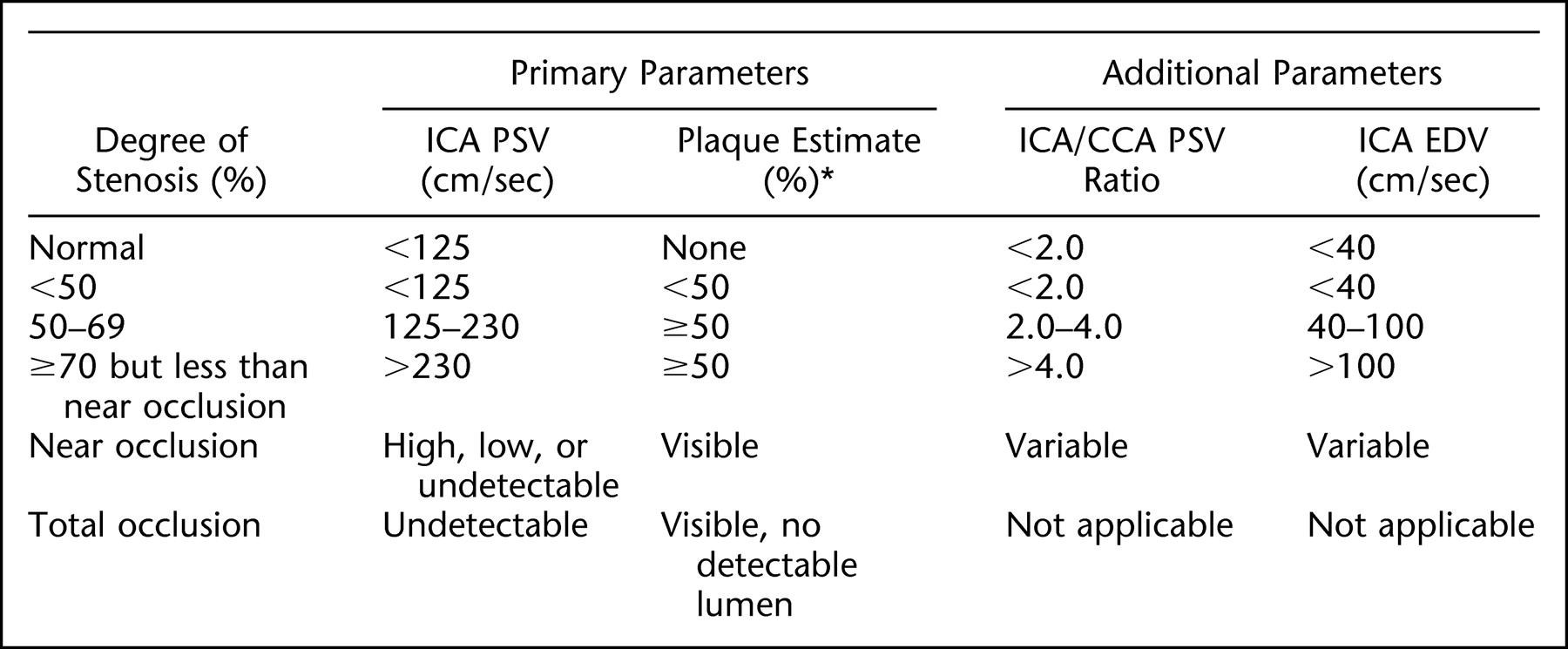
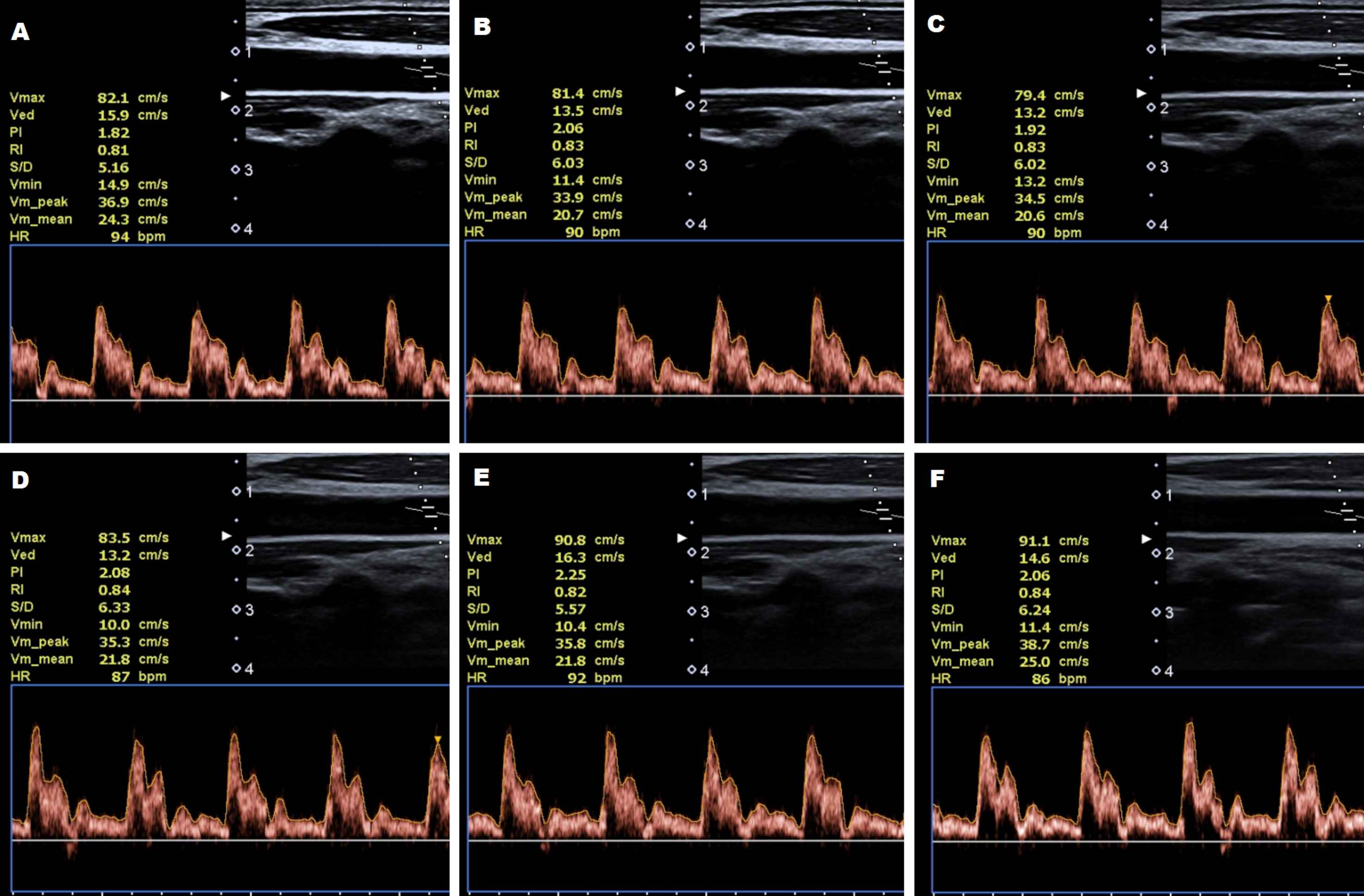
Carotid Velocity Chart - Web a normal ica will have no branches and usually a lower resistance waveform. A complete ultrasound evaluation of the carotid arteries has three components: Web additional criteria refer to the effect of a stenosis on prestenotic flow (common carotid artery), the extent of poststenotic flow disturbances, and derived velocity criteria. Web • velocity increases around a curve • difficult to assign correct doppler angle as direction of blood flow changes rapidly Cca = common carotid artery] normal. Web the maximal peak systolic (ps) velocity is 366 cm/s, the psv index is 6.5 and the st. Web a carotid ultrasound, or carotid duplex, is a painless, safe test that uses sound waves to make images of what your carotid arteries look like on the inside. Web for example, duplex ultrasound studies have consistently shown normal ica/cca peak blood flow velocity ratios of 0.7, 910 and studies of normal angiograms. Web there may be compensatory increased velocity in the contralateral carotid. Web • for ica/cca peak systolic velocity ratio, use the highest psv in the internal carotid artery and the psv in the distal common carotid artery. Web the mean peak systolic velocity in the eca is reported as being 77 cm/sec in normal individuals, and the maximum velocity does not normally exceed 115 cm/sec. Measure the peak systolic (psv) and end diastolic velocities (edv) of the eca. Web in 2021, the intersocietal accreditation commission (iac) introduced modified criteria for carotid duplex interpretation based on peak systolic. Web these recommendations seek to improve quality of care for patients undergoing carotid duplex examinations through standardization of criteria across. Ica end diastolic velocity (edv, cm/s)* mandatory. Web there may be compensatory increased velocity in the contralateral carotid. (1) evaluation of plaque, (2) estimation of ica stenosis. Web internal carotid artery (ica) peak systolic velocity (psv, cm/s) * mandatory. Ica = internal carotid artery; Web additional criteria refer to the effect of a stenosis on prestenotic flow (common carotid artery), the extent of poststenotic flow disturbances, and derived velocity criteria. Web the maximal peak systolic (ps) velocity is 366 cm/s, the psv index is 6.5 and the st. • obtain bilateral brachial blood. Cca = common carotid artery] normal. Ica psv is <125 cm/sec and no plaque or intimal thickening is visible sonographically. Web a normal ica will have no branches and usually a lower resistance waveform. (1) evaluation of plaque, (2) estimation of ica stenosis. Common carotid artery psv (cm/s)*. Web [psv = peak systolic velocity; A complete ultrasound evaluation of the carotid arteries has three components: Web a normal ica will have no branches and usually a lower resistance waveform. • obtain bilateral brachial blood. Web for example, duplex ultrasound studies have consistently shown normal ica/cca peak blood flow velocity ratios of 0.7, 910 and studies of normal angiograms. (1) evaluation of plaque, (2) estimation. Web • velocity increases around a curve • difficult to assign correct doppler angle as direction of blood flow changes rapidly Web the mean peak systolic velocity in the eca is reported as being 77 cm/sec in normal individuals, and the maximum velocity does not normally exceed 115 cm/sec. Web there may be compensatory increased velocity in the contralateral carotid.. (1) evaluation of plaque, (2) estimation of ica stenosis. Carotid duplex ultrasound (cdus) magnetic resonance. • obtain bilateral brachial blood. Ica psv is <125 cm/sec and no plaque or intimal thickening is visible sonographically. Four diagnostic modalities are used to directly image the internal carotid artery: Web a normal ica will have no branches and usually a lower resistance waveform. Cca = common carotid artery] normal. Web for example, duplex ultrasound studies have consistently shown normal ica/cca peak blood flow velocity ratios of 0.7, 910 and studies of normal angiograms. Web the mean peak systolic velocity in the eca is reported as being 77 cm/sec in. Cca = common carotid artery] normal. Common carotid artery psv (cm/s)*. Mary index is 26 mild carotid stenosis. Web a normal ica will have no branches and usually a lower resistance waveform. Four diagnostic modalities are used to directly image the internal carotid artery: Web the mean peak systolic velocity in the eca is reported as being 77 cm/sec in normal individuals, and the maximum velocity does not normally exceed 115 cm/sec. Web internal carotid artery (ica) peak systolic velocity (psv, cm/s) * mandatory. Web additional criteria refer to the effect of a stenosis on prestenotic flow (common carotid artery), the extent of poststenotic. Web • velocity increases around a curve • difficult to assign correct doppler angle as direction of blood flow changes rapidly Web • for ica/cca peak systolic velocity ratio, use the highest psv in the internal carotid artery and the psv in the distal common carotid artery. Web these recommendations seek to improve quality of care for patients undergoing carotid duplex examinations through standardization of criteria across. Web there may be compensatory increased velocity in the contralateral carotid. Ica = internal carotid artery; • obtain bilateral brachial blood. A complete ultrasound evaluation of the carotid arteries has three components: Ica psv is <125 cm/sec and no plaque or intimal thickening is visible sonographically. Web a carotid ultrasound, or carotid duplex, is a painless, safe test that uses sound waves to make images of what your carotid arteries look like on the inside. Mary index is 26 mild carotid stenosis. Four diagnostic modalities are used to directly image the internal carotid artery: Cca = common carotid artery] normal. Web the maximal peak systolic (ps) velocity is 366 cm/s, the psv index is 6.5 and the st. Web additional criteria refer to the effect of a stenosis on prestenotic flow (common carotid artery), the extent of poststenotic flow disturbances, and derived velocity criteria. Common carotid artery psv (cm/s)*. Carotid duplex ultrasound (cdus) magnetic resonance.
Table 1 from Muscle strength and carotid artery flow velocity is
Answers to the Quiz Carotid artery ultrasound on page 76

The internal carotid artery peak systolic velocity to common carotid

Different velocity profiles for the carotid flow pulse as depicted in

Carotid Ultrasound Velocity Chart

Systolic carotid velocity versus percent diameter reduction by

Carotid Ultrasound Velocity Chart

Carotid Ultrasound Velocity Chart

Carotid Ultrasound Velocity Chart
Carotid Doppler Velocity Criteria Chart
Web For Example, Duplex Ultrasound Studies Have Consistently Shown Normal Ica/Cca Peak Blood Flow Velocity Ratios Of 0.7, 910 And Studies Of Normal Angiograms.
Ica End Diastolic Velocity (Edv, Cm/S)* Mandatory.
Web [Psv = Peak Systolic Velocity;
Web Internal Carotid Artery (Ica) Peak Systolic Velocity (Psv, Cm/S) * Mandatory.
Related Post: