Canine Anatomical Chart
Canine Anatomical Chart - Web the following canine anatomy illustrations offer a look at various systems within the dog's body. Although these pictures are fairly basic, they still provide insight that can help the average dog owner gain a working idea of what's beneath all that fur. Web from the bones in their tails to the muscles in their legs, a dog’s body is a marvel of engineering. Web the canine spine is divided into five regions, containing: We discuss the internal and external anatomy of dogs so that you can see that, despite individual differences, there is a reason they are all considered part of the same species. Images are available in 3 different planes (transverse, sagittal and dorsal), with two kind of contrast (bone and soft tissues). A dog’s neck and shoulders. Some of the different canine joints are labeled. Web dog anatomy comprises the anatomical study of the visible parts of the body of a domestic dog. A brief outline of diagnostic, Web the anatomy of a dog includes its skeletal structure, reproductive system, the internal organs, and its external appearance. Cannes film festival (un certain regard) cast: Feb 24, 2022 | last update: Web the canine spine is divided into five regions, containing: • the dorsal plane divides the dog into ventral and dorsal portions. Like all teeth, canines have a crown (the part above the gum), a neck and a root (the part inside the bone). If this plane were in the midline of the body, this is the median plane or median sagittal plane. Have you ever wondered how your dog’s body works? Let’s learn about your dog's anatomy to help you get. Web dog anatomy comprises the anatomical study of the visible parts of the body of a domestic dog. Images are available in 3 different planes (transverse, sagittal and dorsal), with two kind of contrast (bone and soft tissues). The detailing of these structures changes based on dog breed due to the huge variation of size in dog breeds. Posterior view. Web from the bones in their tails to the muscles in their legs, a dog’s body is a marvel of engineering. Let’s learn about your dog's anatomy to help you get better acquainted with your pup’s body parts, from head to tail! Feb 24, 2022 | last update: Web dog anatomy comprises the anatomical study of the visible parts of. Feb 24, 2022 | last update: Web this is one of our bestselling veterinary charts in the canine anatomy series, which includes the canine muscular system and canine skeletal anatomy charts. Here are presented scientific illustrations of the canine muscles and skeleton from different anatomical standard views (lateral, medial, cranial, caudal, dorsal, ventral / palmar.). Web dog anatomy comprises the. Understand the musculoskeletal, nervous and internal organ systems easily with these wall hangings in lamination or paper. Seven cervical vertebrae, thirteen thoracic vertebrae, seven lumbar vertebrae and three sacral vertebrae. Posterior view of the skeleton with lateral view of the skull. We discuss the internal and external anatomy of dogs so that you can see that, despite individual differences, there. Some of the different canine joints are labeled. Dog simulators are available for vet surgical training. In this article, you will learn the location of different organs from the different systems (like skeletal, digestive, respiratory, urinary, cardiovascular, endocrine, nervous, and special sense) of a dog with their important anatomical features. Songs are ranked by peak position. Anatomical terms you should. The canine tail is an extension of the spine. Dog simulators are available for vet surgical training. This article will explore the fascinating world of dog anatomy, providing insight into the unique physical attributes that make man’s best friend such a remarkable and resilient animal. • the dorsal plane divides the dog into ventral and dorsal portions. Web the following. This article will explore the fascinating world of dog anatomy, providing insight into the unique physical attributes that make man’s best friend such a remarkable and resilient animal. Web anatomy of the canine whole body on ct. Web our veterinary anatomy posters and anatomical charts are scientifically accurate. In this article, you will learn the location of different organs from. Muscle, organ and skeletal anatomy). Web dog anatomy details the various structures of canines (e.g. Although these pictures are fairly basic, they still provide insight that can help the average dog owner gain a working idea of what's beneath all that fur. Cannes film festival (un certain regard) cast: Anatomical terms you should know. Web here are presented scientific illustrations of the canine skeleton, containing the main joints of the dog and its structures from different standard anatomical views (cranial, caudal, lateral, medial, dorsal, palmar.). This article will explore the fascinating world of dog anatomy, providing insight into the unique physical attributes that make man’s best friend such a remarkable and resilient animal. The size of the tail, and therefore the number of bones in a dog, is dependent on the length of their tail. Here are presented scientific illustrations of the canine muscles and skeleton from different anatomical standard views (lateral, medial, cranial, caudal, dorsal, ventral / palmar.). Positional and directional terms, general terminology and anatomical orientation are also illustrated. Fully labeled illustrations and diagrams of the dog (skeleton, bones, muscles, joints, viscera, respiratory system, cardiovascular system). The following paragraphs explain all these aspects in brief, along with diagrams, which will help you understand them better. Choose from a large selection of topics on canine, feline, equine, and bovine anatomy. The detailing of these structures changes based on dog breed due to the huge variation of size in dog breeds. Web the canine spine is divided into five regions, containing: Web this is one of our bestselling veterinary charts in the canine anatomy series, which includes the canine muscular system and canine skeletal anatomy charts. • the sagittal plane divides the dog into right and left portions. Web anatomy of the canine whole body on ct. Web humans have four canine teeth, two maxillary canine teeth (left and right) and two mandibular canine teeth (left and right). If this plane were in the midline of the body, this is the median plane or median sagittal plane. The outer surface is a thin layer of enamel which covers the inner dentin.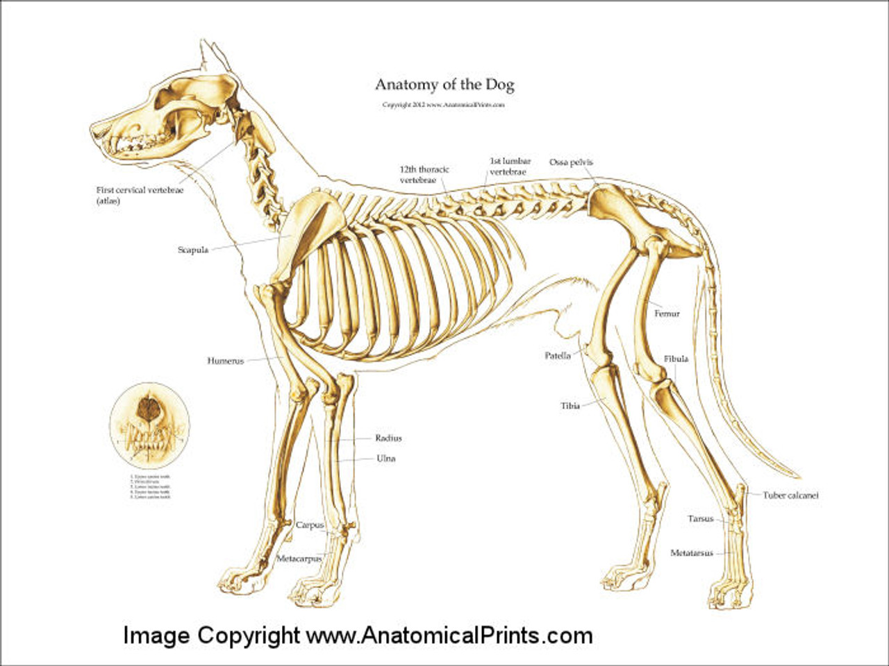
Canine Skeleton Poster Clinical Charts and Supplies
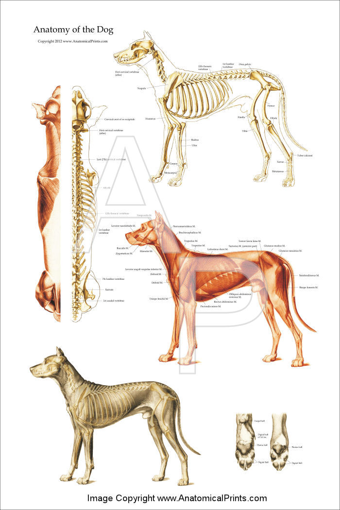
Dog Anatomical Chart Bones and Muscles
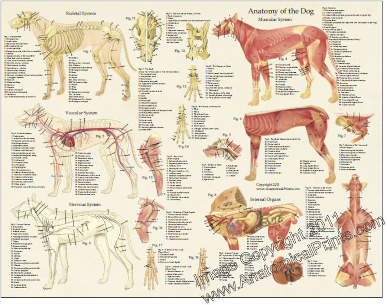
Dog Anatomy Laminated Poster Clinical Charts and Supplies

the anatomy of a dog's body and its major skeletal systems are shown in
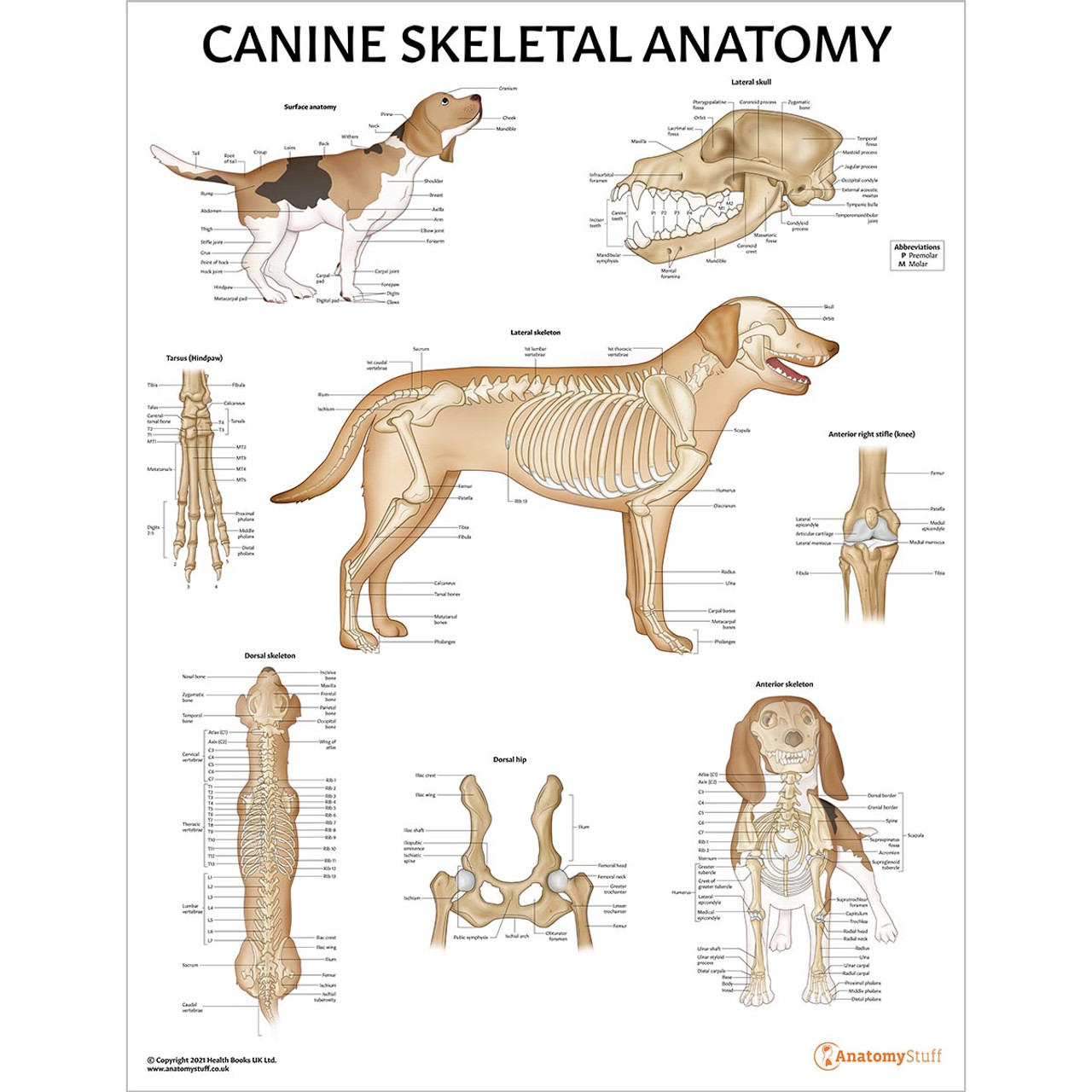
Canine Skeletal Anatomy Laminated Chart Dog Skeleton Poster

Canine Anatomy, Complete Set of 3 Charts. Buy The Set and Save! Amazon
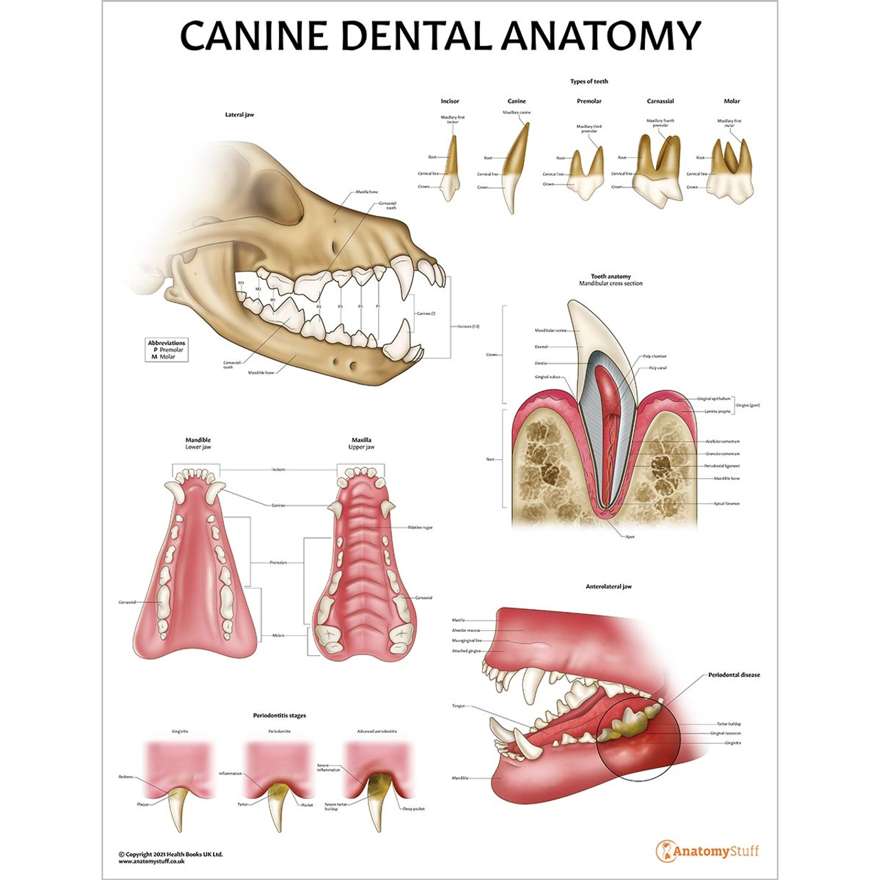
Canine Anatomy Models, Charts & Simulators Dog Skeleton Page 3
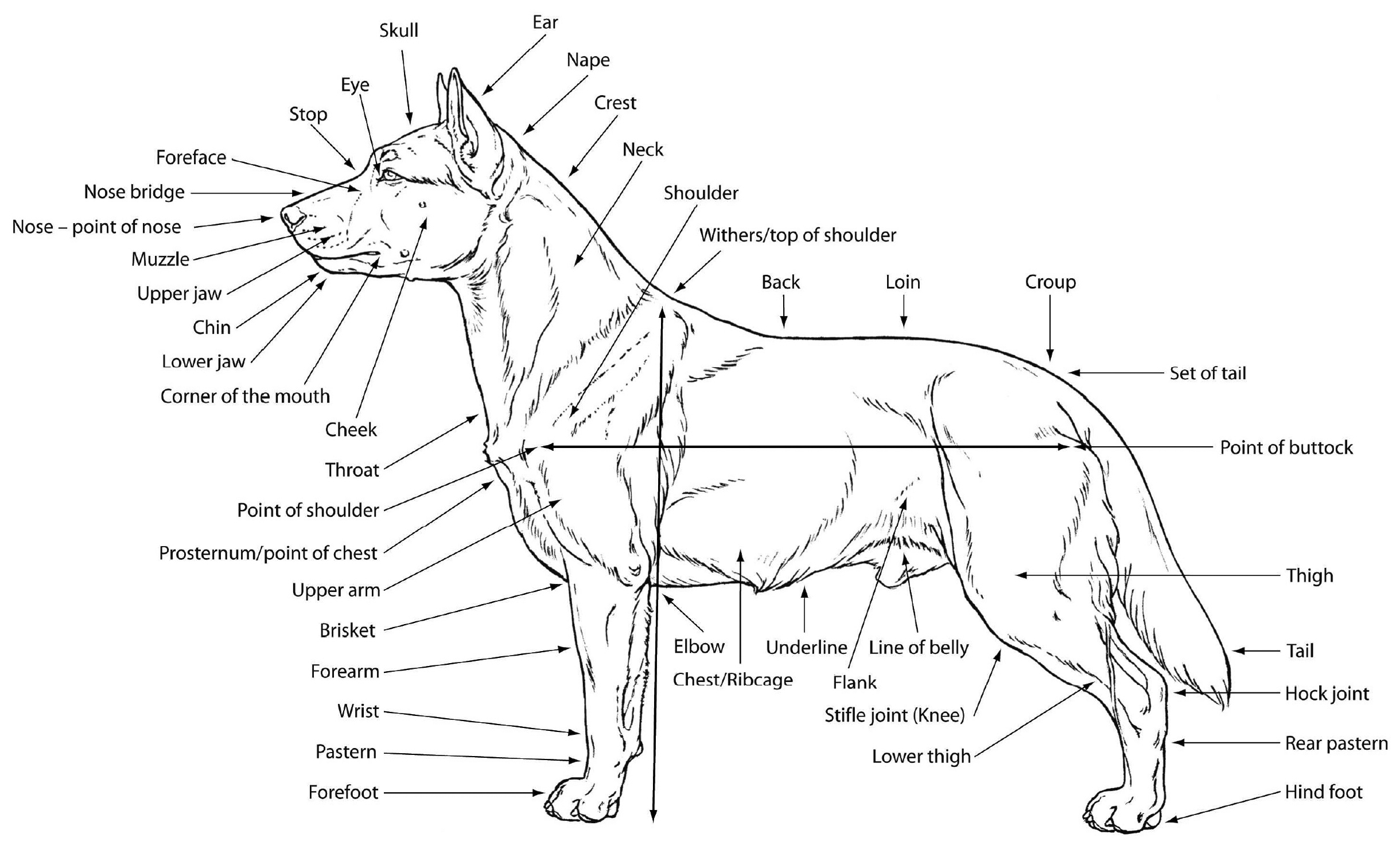
M. Douglas Wray Dog Anatomy

Canine Internal Anatomy Chart Poster Laminated ubicaciondepersonas
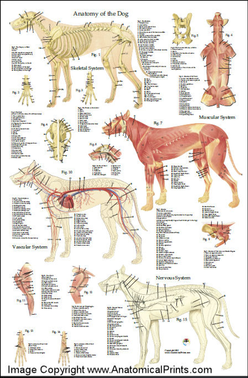
Canine Internal Anatomy Chart Poster Laminated ubicaciondepersonas
• The Dorsal Plane Divides The Dog Into Ventral And Dorsal Portions.
Web The Anatomy Of A Dog Includes Its Skeletal Structure, Reproductive System, The Internal Organs, And Its External Appearance.
Although These Pictures Are Fairly Basic, They Still Provide Insight That Can Help The Average Dog Owner Gain A Working Idea Of What's Beneath All That Fur.
Dog Simulators Are Available For Vet Surgical Training.
Related Post: