Anatomy Of The Teeth Anatomical Chart
Anatomy Of The Teeth Anatomical Chart - Web most adults have 32 permanent teeth, including eight incisors, four canines, eight premolars and 12 molars. Watch the video tutorial now. They cut and crush foods, making them easier to swallow. The large central image shows a detailed cross section of a tooth and surrounding gum and bone with clearly labeled anatomic features. Each tooth has a crown and a root. Web the anatomy of a tooth divides into two main sections: Your teeth play a big role in digestion. Prefer to learn by doing? The two types are the central incisors and lateral incisors. Web we’ll go over the anatomy of a tooth and the function of each part. Brightly colored, user friendly chart covering the anatomy of the teeth. Web 4.1 20 ratings. Use our diagram to learn more about teeth numbers and placement. Web the anatomy of a tooth divides into two main sections: Fully labeled illustrations of the teeth with dental terminology (orientation, surfaces, cusps, roots numbering systems) and detailed images of each permanent tooth. The large central image shows a detailed cross section of a tooth and surrounding gum and bone with clearly labeled anatomic features. Function and types of teeth. Structure and surrounding structures of a tooth seen in cross section. There are dental charts showing disorders of the jaw and other diseases of the dental structure. (the function of teeth as they. Web dental anatomy is a field of anatomy dedicated to the study of human tooth structures. Brightly colored, user friendly chart covering the anatomy of the teeth. Watch the video tutorial now. The large central image shows a detailed cross section of a tooth and surrounding gum and bone with clearly labeled anatomic features. Incisors and canines can be classified. Web anatomy of the teeth anatomical chart company staff,f. It's nicole from kenhub here, and welcome to our tutorial on the anatomy of the tooth. Web in the mouth, the bone holding the bottom row of teeth is the mandible, and the bone holding the top row of teeth is the maxilla. Web brightly colored, user friendly chart covering the. The crown of the tooth is what is visible in the oral cavity, and the root of the tooth is embedded into the bony ridge of the upper and lower jaws called the alveolar process via attachment to the periodontal ligament. In this page, we are going to study each one of the above types, learn how they are numbered,. Get dental skull and jaw models at the best possible prices. The large central image shows a detailed cross section of a tooth and surrounding gum and bone with clearly labeled anatomic features. Web the anatomy of a tooth divides into two main sections: It's nicole from kenhub here, and welcome to our tutorial on the anatomy of the tooth.. The large central image shows a detailed cross section of a tooth and surrounding gum and bone with clearly labeled anatomic features. Each tooth has a crown and a root. Premolars are only present in the permanent dentition. It's nicole from kenhub here, and welcome to our tutorial on the anatomy of the tooth. The maxillary (upper) arch and the. They cut and crush foods, making them easier to swallow. Web the anatomy of a tooth divides into two main sections: The development, appearance, and classification of teeth fall within its purview. The large central image shows a detailed cross section of a tooth and surrounding gum and bone with clearly labeled anatomic features. So, thanks for joining me in. Web atlas of dental anatomy: The initial deciduous (primary) teeth and the successive permanent (secondary) teeth. This leaves up to eight adult teeth in each quadrant and separates the opposing pairs within the same alveolar bone as well as their counterparts in the opposing jaw. Web we have detailed dental anatomical models for dentistry and oral care. Web brightly colored,. Web atlas of dental anatomy: The two types are the central incisors and lateral incisors. The large central image shows a detailed cross section of a tooth and surrounding gum and bone with clearly labeled anatomic features. Also includes labeled illustrations of the following: The crown and the root. Watch the video tutorial now. The two types are the central incisors and lateral incisors. Premolars are only present in the permanent dentition. Web we’ll go over the anatomy of a tooth and the function of each part. Web most adults have 32 permanent teeth, including eight incisors, four canines, eight premolars and 12 molars. There are typically 20 deciduous teeth divided evenly across the maxilla and mandible. Structure and surrounding structures of a tooth seen in cross section. Incisors and canines can be classified further as anterior teeth and molars and premolars as posterior teeth. Brightly colored, user friendly chart covering the anatomy of the teeth. Teeth are positioned in alveolar sockets and connected to the bone by a suspensory periodontal ligament. Use our diagram to learn more about teeth numbers and placement. Web the anatomy of a tooth divides into two main sections: Temporomandibular joint posters and much more are also available. There are 8 incisors in both the permanent and primary dentition, with four in each dental arch. Web the anterior teeth are the twelve teeth in the front of the mouth, while the posterior teeth are the teeth in the back of the mouth. The large central image shows a detailed cross section of a tooth and surrounding gum and bone with clearly labeled anatomic features.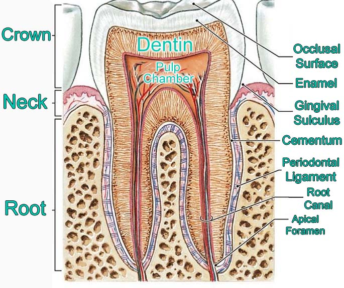
Anatomy Of Tooth Anatomy Book
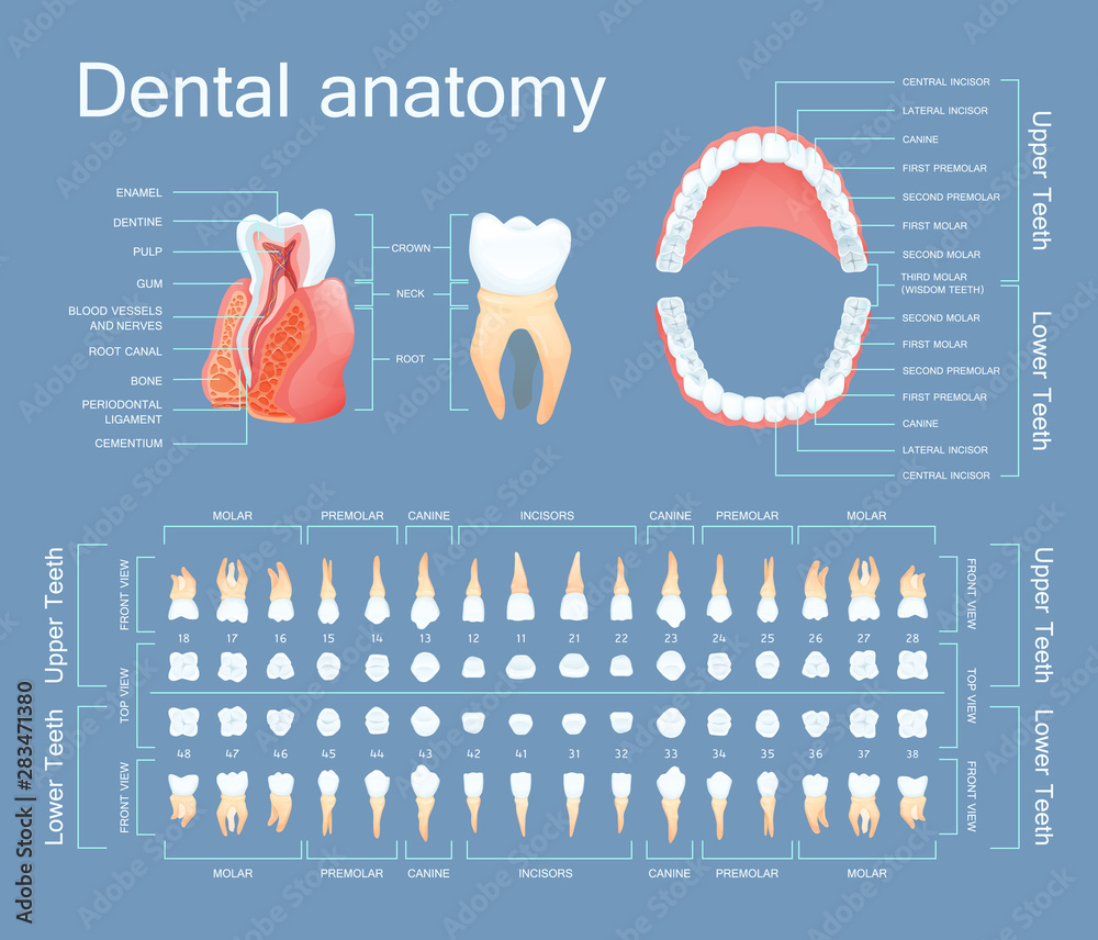
Human dental anatomy. Tooth anatomy numbering infographics. Sectional
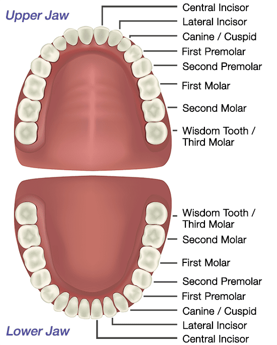
The Anatomy of Your Teeth Sabka Dentist Top Dental Clinic Chain In
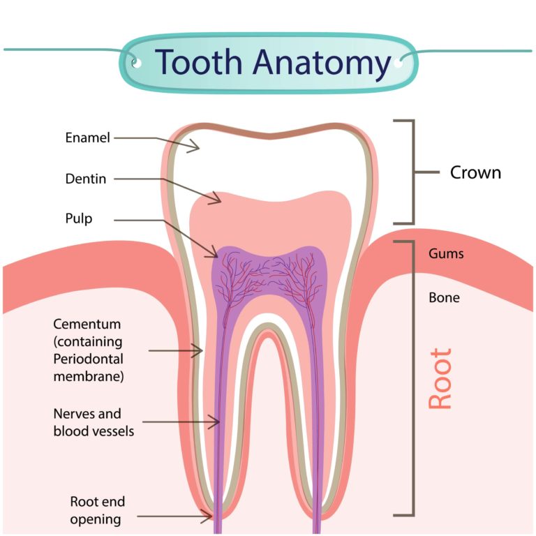
The Anatomy of Your Teeth

Anatomy of the Teeth Anatomical Chart Poster (20" x 26") NEW anatomy
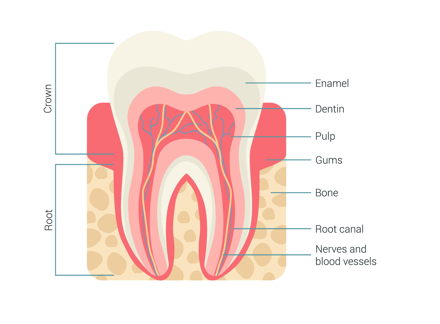
Tooth Anatomy Milford Family Dentistry

The Different Types of Teeth Gentle Dentist
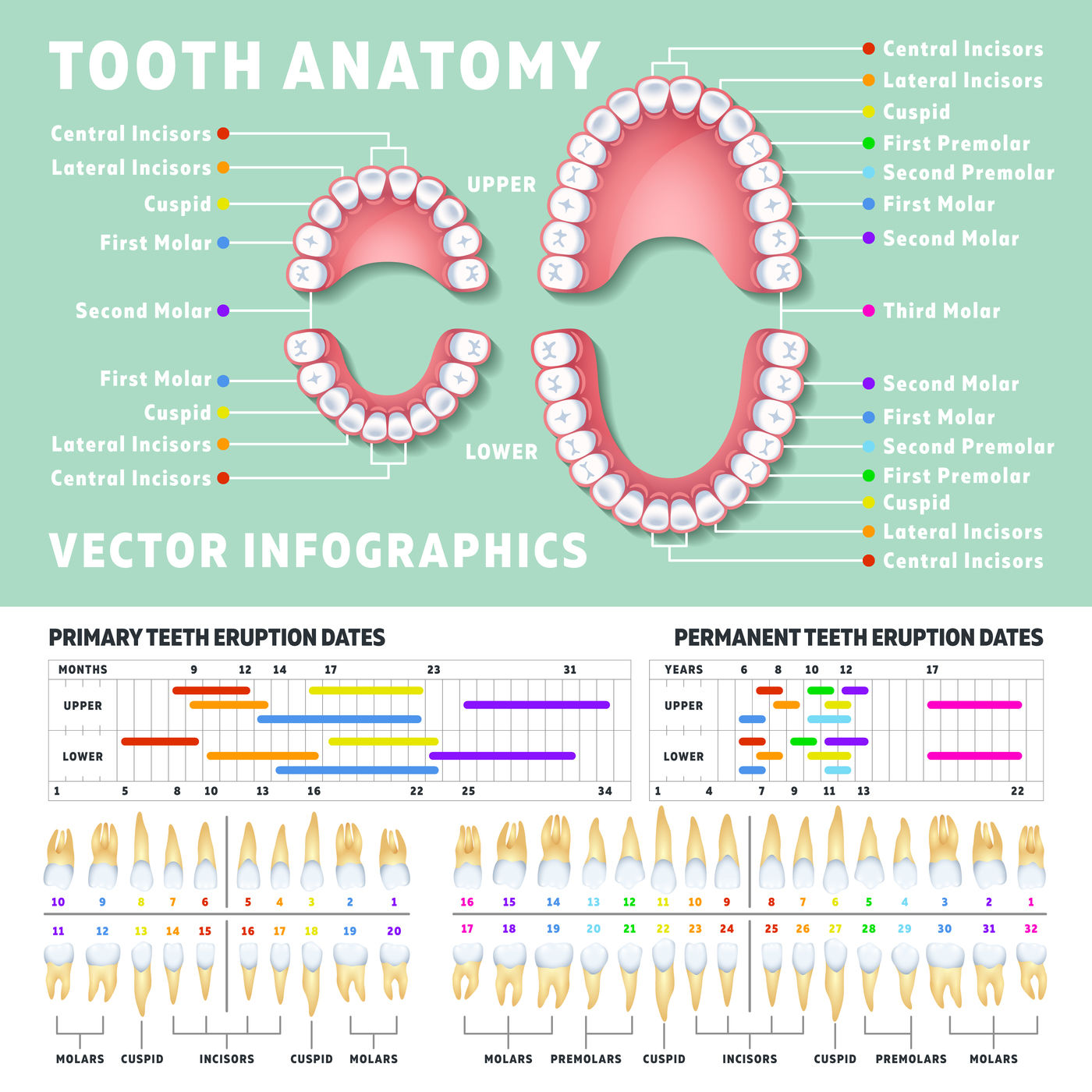
Orthodontist human tooth anatomy vector infographics with teeth diagra
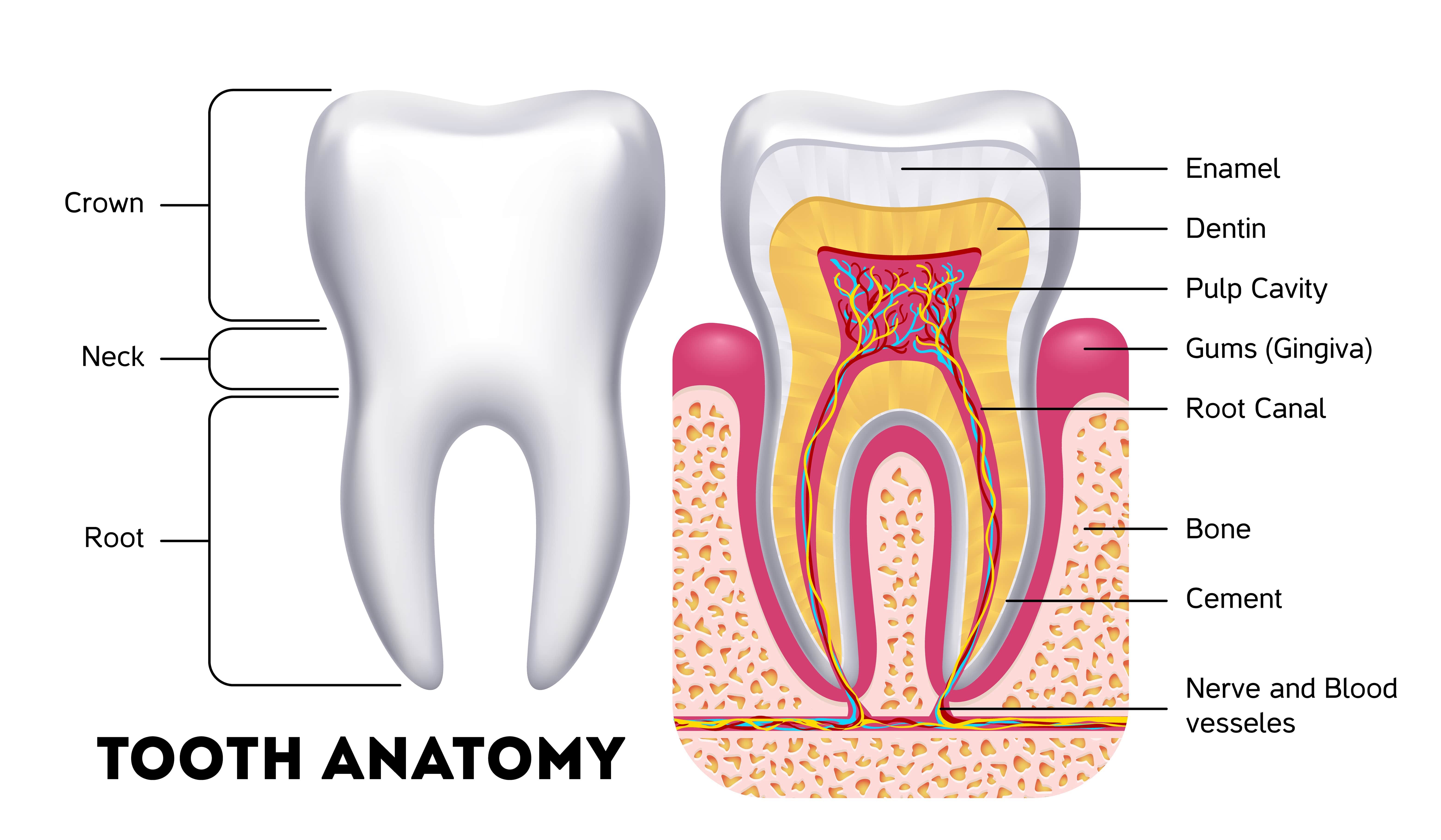
Anatomy Of The Teeth Anatomical Chart Poster Prints Images and Photos

Anatomy of The Teeth Anatomical Chart 20'' x 26''
The Initial Deciduous (Primary) Teeth And The Successive Permanent (Secondary) Teeth.
Web Anatomy Of The Teeth Anatomical Chart Company Staff,F.
The Large Central Image Shows A Detailed Cross Section Of A Tooth And Surrounding Gum And Bone With Clearly Labeled Anatomic Features.
Brightly Colored, User Friendly Chart Covering The Anatomy Of The Teeth.
Related Post: